 OpenDissect
All fractures › Dislocations
Shoulder dislocation
GlenohumeralAnterior, posterior, inferior · Hill–Sachs, Bankart, greater tuberosity
See it in 3D on the real bones, displace and reduce it, with simulated X-ray and CT — free, in your browser.
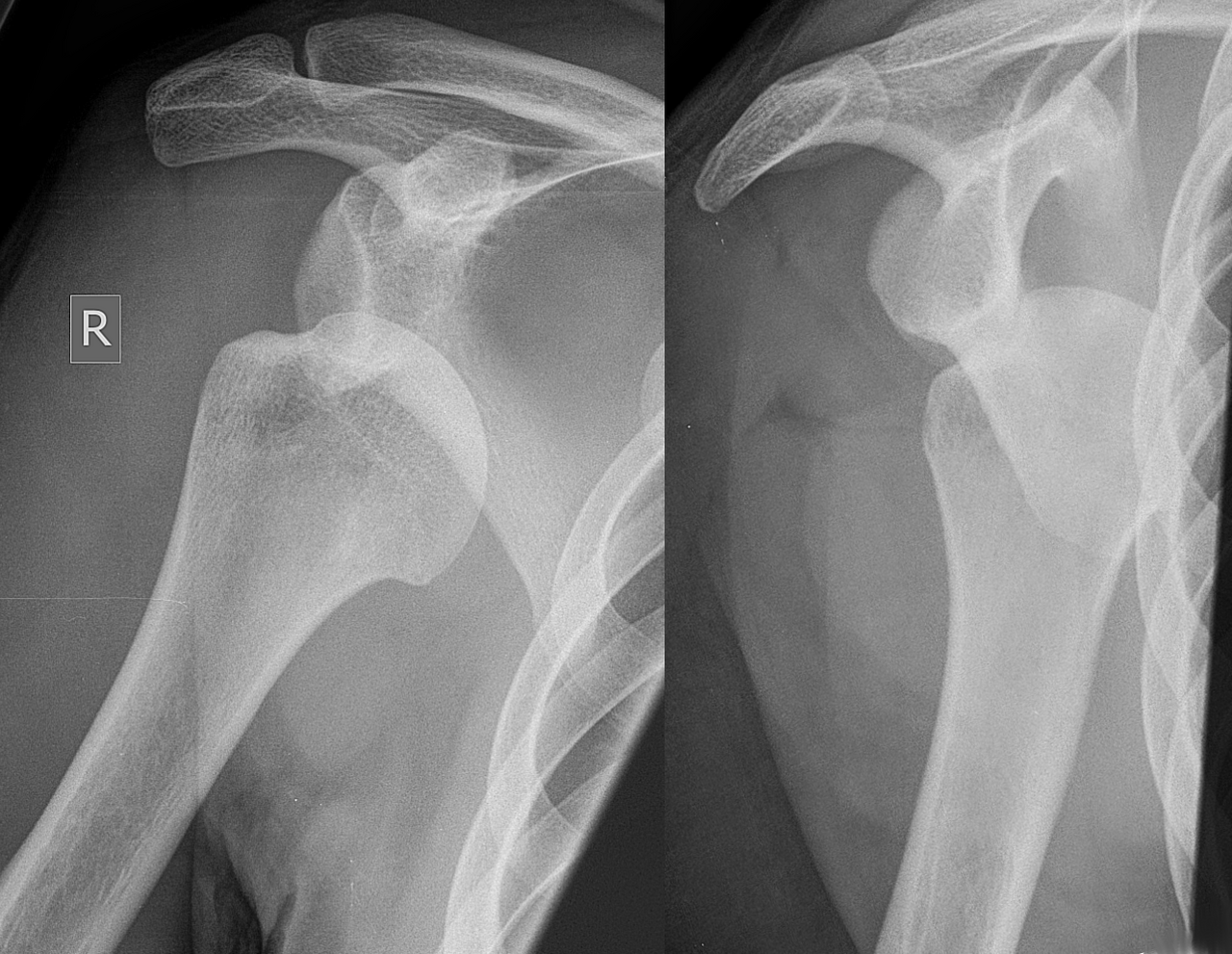
Open in 3D →The glenohumeral joint is the most often dislocated large joint: a big head on a small, shallow glenoid, held mainly by soft tissue. Most dislocations are anterior. The young re-dislocate (labrum torn off: Bankart), the older tear the rotator cuff or break the greater tuberosity.
Mechanism
Anterior: fall or tackle with the arm abducted and externally rotated. Posterior: seizure, electric shock, fall on the outstretched arm in internal rotation. Inferior: hyperabduction.
Clinical findings
- Squared-off shoulder, arm held away from the body, cannot rotate.
- Before and after reduction: axillary nerve (sensation over the regimental badge area, deltoid contraction), radial pulse and hand neurology.
- Over about 40: test the rotator cuff once the pain settles.
- Posterior dislocation: arm locked in internal rotation, cannot externally rotate. Easily missed.
Imaging
- AP in the scapular plane plus a Y view or axillary view: always two planes, before and after reduction.
- Look for a greater tuberosity or glenoid rim fracture, and a fracture of the humeral neck (do not try to reduce a fracture-dislocation in the emergency room).
- After a first dislocation in the young or with a large bony lesion: CT; MRI arthrogram for labrum and cuff.
Classification
- Direction
- Anterior (subcoracoid, subglenoid, subclavicular), posterior, inferior (luxatio erecta).
- Hill–Sachs
- Posterolateral humeral head impaction from the glenoid rim.
- Bankart
- Anteroinferior labrum (soft) or rim fracture (bony) torn off the glenoid.
- Fracture-dislocation
- With greater tuberosity, surgical neck or anatomical neck fracture (see proximal humerus).
Types shown in 3D
- Anterior (subcoracoid)
- About 95% of shoulder dislocations. The head comes out in front of the glenoid and comes to lie under the coracoid; the arm is held slightly abducted and externally rotated, the shoulder looks squared off.
- Posterior
- Rare (2–4%) and often missed: after epileptic seizures and electric shocks. The arm is locked in internal rotation; the AP view shows the "lightbulb" head and an empty anterior glenoid. Always get an axillary or Y view.
- Inferior (luxatio erecta)
- The head is levered below the glenoid with the arm locked straight overhead. High rate of axillary nerve and artery injury and of rotator cuff tears or greater tuberosity fractures.
- Anterior + greater tuberosity
- In patients over about 40 the cuff often pulls off the greater tuberosity instead of tearing. The fragment usually reduces with the head; it needs fixation if it stays displaced more than about 5 mm.
- Anterior + bony Bankart
- The head shears off the anterior-inferior rim of the glenoid. A large rim fragment (more than about 20% of the glenoid width) leaves the shoulder unstable: fix it or reconstruct the glenoid (Latarjet).
Treatment
- Reduction
- Analgesia/sedation, then a gentle technique: external rotation (Kocher’s first step only), Cunningham massage, FARES or traction–countertraction; Hippocratic method only gently. Check neurovascular status and repeat the X-ray.
- After a first dislocation
- Sling for comfort 1–3 weeks, then physiotherapy. Young active patients (high recurrence) or a large bony Bankart: consider early arthroscopic Bankart repair or Latarjet.
- Fracture-dislocation
- Displaced greater tuberosity (> 5 mm): fixation. Neck fracture with dislocation: reduction in theatre and fixation or arthroplasty in the elderly.
- Posterior / chronic
- Closed reduction under anaesthesia; large reverse Hill–Sachs lesions need McLaughlin procedure, grafting or arthroplasty.
Aftercare
Physiotherapy for rotator cuff and scapular control; avoid abduction–external rotation for 6 weeks after a first anterior dislocation.
Complications
- Recurrence (up to 80–90% under 20 years old)
- Axillary nerve palsy
- Rotator cuff tear (older patients)
- Missed posterior dislocation
- Fracture during reduction (iatrogenic neck fracture)
See it in 3D on the real bones, displace and reduce it, with simulated X-ray and CT — free, in your browser.
Open in 3D →