 OpenDissect
All fractures › Dislocations
Knee dislocation
Schenck KD I–VMultiligament injury · popliteal artery
See it in 3D on the real bones, displace and reduce it, with simulated X-ray and CT — free, in your browser.
Open in 3D →A tibiofemoral dislocation tears at least two of the four main ligaments and threatens the popliteal artery, which is fixed above (adductor hiatus) and below (soleal arch). Many reduce spontaneously before arrival, so think of it in any multiligament injury.
Mechanism
High energy (road traffic) or low energy in obese patients (stepping off a kerb). Hyperextension causes anterior, a dashboard blow posterior dislocation.
Clinical findings
- Reduce immediately and recheck pulses.
- Ankle–brachial index in every case: below 0.9 means CT angiography; a pulseless limb goes to theatre.
- Common fibular nerve (foot drop) in up to a quarter. Compartment syndrome.
Imaging
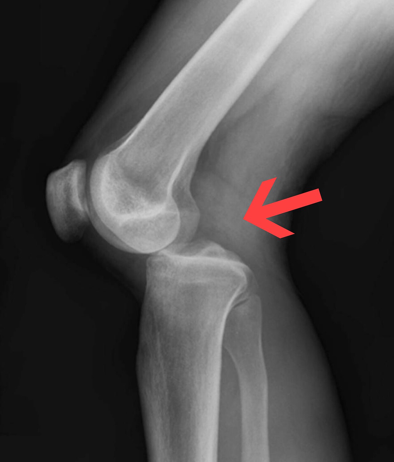
- AP and lateral (look for Segond fracture, tibial spine or fibular head avulsion). MRI for the ligaments once the limb is safe.
Classification
- Kennedy
- By direction: anterior, posterior, medial, lateral, rotatory.
- Schenck KD
- I one cruciate + collateral, II both cruciates, III both cruciates + MCL (M) or posterolateral corner (L), IV all four, V with fracture; C vascular, N nerve injury.
Types shown in 3D
- Anterior
- The most common direction, from hyperextension. The popliteal artery is tethered above and below the knee and tears or thromboses: check pulses and the ankle–brachial index in every case; CT angiography if abnormal.
- Posterior
- Dashboard blow to the front of the tibia. Same vascular risk; the common fibular nerve is often stretched too.
- Lateral
- Valgus force: the tibia goes out sideways, with the medial collateral and both cruciates torn.
Treatment
- Emergency
- Reduction, splint in 20° flexion or spanning external fixator if unstable, vascular repair if needed (with fasciotomies).
- Ligaments
- Staged or early (within 3 weeks) reconstruction of the cruciates and repair/reconstruction of the collaterals and posterolateral corner in most active patients.
Aftercare
Long rehabilitation; stiffness and residual laxity are common.
Complications
- Popliteal artery injury (amputation if missed)
- Common fibular nerve palsy
- Stiffness / arthrofibrosis
- Residual instability
- Post-traumatic arthritis
See it in 3D on the real bones, displace and reduce it, with simulated X-ray and CT — free, in your browser.
Open in 3D →