 OpenDissect
All fractures › Dislocations
Ankle fracture-dislocation
AO/OTA 44B3 / 44CTrimalleolar, bimalleolar, pure dislocation
See it in 3D on the real bones, displace and reduce it, with simulated X-ray and CT — free, in your browser.
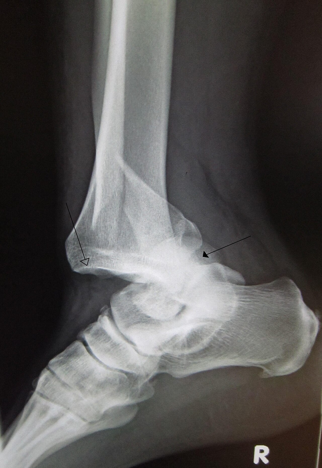
Open in 3D →When the malleoli or the ligaments that hold the talus fail together, the talus leaves the mortise. The skin over the medial malleolus is stretched white and will necrose within hours: reduction comes before anything else.
Mechanism
Supination–external rotation or pronation forces with high energy, falls and sport; often in elderly patients with osteoporotic bone.
Clinical findings
- Obvious deformity; skin blanched over the medial side. Check pulses (dorsalis pedis, posterior tibial) and sensation.
- Reduce and splint immediately, then X-ray. Open fracture-dislocations: antibiotics, tetanus, wound cover.
Imaging
- AP mortise and lateral after reduction: medial clear space, talar position, posterior malleolus size. CT for the posterior malleolus and joint impaction.
Classification
- Weber / AO 44
- A infra-, B trans-, C suprasyndesmotic fibula fracture (see the ankle fracture entries).
- Lauge-Hansen
- Mechanism: supination–external rotation, pronation–external rotation, supination–adduction, pronation–abduction.
Types shown in 3D
- Trimalleolar fracture-dislocation
- Lateral, medial and posterior malleoli broken; the talus dislocates posterolaterally out of the mortise and tents the skin. Reduce and splint immediately in the emergency room, before any X-ray if the skin is at risk; then fix (often after an external fixator).
- Bimalleolar fracture-dislocation
- Fibula and medial malleolus broken, the talus shifts laterally with the fibula. Any talar shift means an unstable ankle: fix.
- Pure ankle dislocation
- Rare: the talus leaves the mortise without a fracture (high energy, often open). Reduce urgently; usually treated with a cast or fixator, ligaments repaired when needed.
Treatment
- Immediately
- Closed reduction (traction and correction of the deformity), well-padded backslab, elevation.
- Definitive
- ORIF of the fibula, medial malleolus and a significant posterior malleolus; syndesmosis fixation if unstable. Severe swelling or open injury: spanning external fixator first, fixation when the skin allows. Fragile elderly: hindfoot nail.
Aftercare
Protected weight bearing about 6 weeks.
Complications
- Skin necrosis and infection
- Malreduction (syndesmosis)
- Post-traumatic arthritis
- Stiffness, CRPS
See it in 3D on the real bones, displace and reduce it, with simulated X-ray and CT — free, in your browser.
Open in 3D →