 OpenDissect
All fractures › Dislocations
Hip dislocation
Thompson–Epstein · PipkinPosterior, anterior, with wall or head fracture
See it in 3D on the real bones, displace and reduce it, with simulated X-ray and CT — free, in your browser.
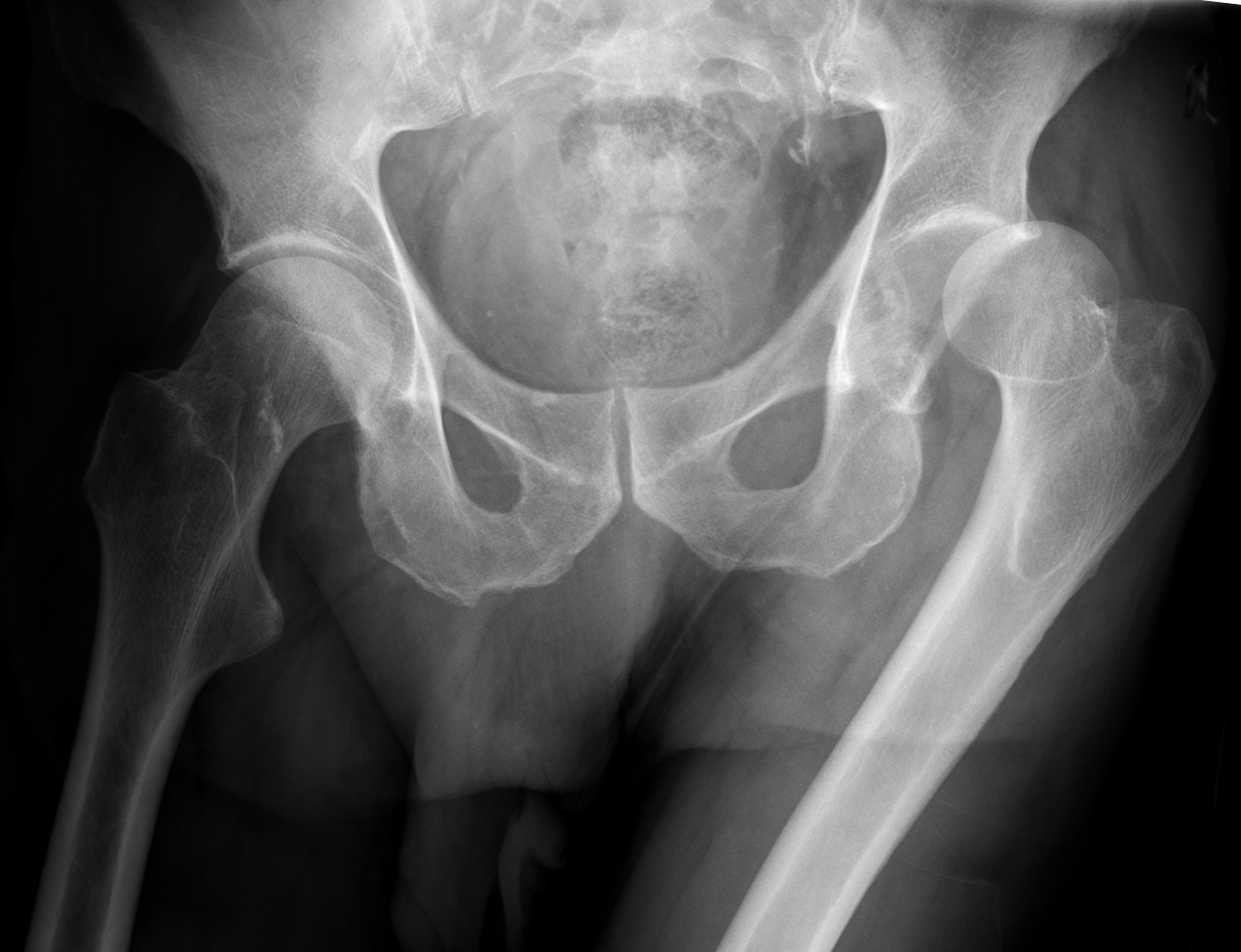
Open in 3D →A high-energy injury: it takes a lot to push the femoral head out of its deep socket. Most dislocations are posterior, often with a posterior wall or femoral head fracture. It is an emergency: the longer the head stays out, the higher the risk of avascular necrosis.
Mechanism
Posterior: dashboard injury, force along the femur with the hip and knee flexed. Anterior: forced abduction and external rotation.
Clinical findings
- ATLS first (associated injuries are the rule).
- Posterior: leg short, flexed, adducted and internally rotated. Anterior: abducted and externally rotated.
- Sciatic nerve (mainly fibular division: foot dorsiflexion) before and after reduction; examine the knee (patella, PCL).
Imaging
- AP pelvis: the femoral heads should look equal in size; Shenton line broken. CT after reduction for loose fragments in the joint, wall and head fractures.
Classification
- Thompson–Epstein (posterior)
- I simple or minor fragment, II large posterior wall, III comminuted wall, IV acetabular floor, V femoral head.
- Pipkin (femoral head)
- I below the fovea, II above the fovea, III with neck fracture, IV with acetabular fracture.
- Epstein (anterior)
- Superior (pubic) or inferior (obturator) type.
Types shown in 3D
- Posterior
- About 90%. Dashboard injury: force along the femur with the hip flexed and adducted. The leg is short, flexed, adducted and internally rotated. Reduce within 6 hours to protect the head’s blood supply.
- Posterior + posterior wall
- Thompson–Epstein II–III: the head breaks off the posterior wall of the acetabulum. A large or comminuted wall fragment leaves the hip unstable after reduction: fixation (Kocher–Langenbeck).
- Pipkin (femoral head)
- A shear fragment of the femoral head stays in the acetabulum while the rest dislocates. Pipkin I below the fovea (outside weight bearing), II above it, III with a neck fracture, IV with an acetabular fracture.
- Anterior
- About 10%: forced abduction and external rotation. The head lies over the obturator foramen (inferior) or the pubis (superior); the leg is abducted and externally rotated. Watch the femoral vessels and nerve.
Treatment
- Emergency reduction
- Within 6 hours, under sedation or general anaesthesia with relaxation (Allis or Bigelow technique). Never force it: a neck fracture must be excluded first.
- After reduction
- CT. Stable concentric hip without fragments: mobilise with protected weight bearing. Incarcerated fragments, unstable posterior wall or displaced head fracture: open reduction and fixation (Kocher–Langenbeck or surgical hip dislocation).
Aftercare
Protected weight bearing 4–6 weeks; MRI at about 3 months if pain suggests avascular necrosis.
Complications
- Avascular necrosis of the femoral head
- Post-traumatic arthritis
- Sciatic nerve palsy
- Recurrent dislocation (rare)
- Heterotopic ossification
See it in 3D on the real bones, displace and reduce it, with simulated X-ray and CT — free, in your browser.
Open in 3D →