 OpenDissect
All fractures › Dislocations
Patellar dislocation
LateralMPFL tear · osteochondral fracture
See it in 3D on the real bones, displace and reduce it, with simulated X-ray and CT — free, in your browser.
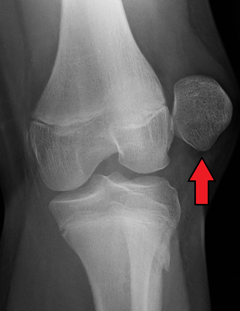
Open in 3D →The patella slips out of the trochlear groove, almost always laterally, tearing the medial patellofemoral ligament (MPFL). Typical in teenagers and young adults, especially with a shallow trochlea or a high patella.
Mechanism
Twisting with the foot planted and the knee slightly flexed and in valgus (sport, dance). Less often a direct blow.
Clinical findings
- The patella sits on the lateral side of the knee, knee held flexed; it often reduces as the knee is straightened.
- Haemarthrosis suggests an osteochondral fracture. Medial tenderness at the adductor tubercle (MPFL). Apprehension test later.
Imaging
- AP, lateral and skyline (Merchant) views: look for an osteochondral fragment. MRI after a first dislocation with haemarthrosis, and to assess trochlear dysplasia and patellar height.
Classification
- First-time vs recurrent
- Risk factors
- Trochlear dysplasia (Dejour), patella alta (Caton–Deschamps), TT–TG distance > 20 mm, valgus, femoral anteversion, laxity.
Types shown in 3D
- Lateral dislocation
- The patella almost always goes laterally, with the knee slightly flexed and the leg twisting inwards over a planted foot. The medial patellofemoral ligament (MPFL) tears. Often reduces when the knee is straightened.
- With osteochondral fracture
- As the patella relocates it strikes the lateral femoral condyle: an osteochondral fragment from the medial facet (or the condyle) can float free in the joint. A large fragment is fixed back, a small one removed.
Treatment
- First dislocation
- Reduce by extending the knee, brace for comfort, physiotherapy (quadriceps and hip control).
- Osteochondral fracture
- Arthroscopic or open fixation of a large fragment, removal of a small one, usually with MPFL repair.
- Recurrent
- MPFL reconstruction, plus tibial tubercle osteotomy or trochleoplasty for marked anatomical risk factors.
Aftercare
Return to sport after 3–6 months when strength is restored.
Complications
- Recurrence (about 1 in 3 after a first dislocation)
- Patellofemoral pain and arthritis
- Loose bodies
See it in 3D on the real bones, displace and reduce it, with simulated X-ray and CT — free, in your browser.
Open in 3D →