 OpenDissect
All fractures › Dislocations
Acromioclavicular dislocation
Rockwood I–VIAC and coracoclavicular ligaments
See it in 3D on the real bones, displace and reduce it, with simulated X-ray and CT — free, in your browser.
Open in 3D →A fall onto the point of the shoulder drives the acromion down while the clavicle stays: the acromioclavicular ligaments tear first, then the coracoclavicular ligaments (conoid and trapezoid). The more ligaments torn, the higher the lateral clavicle stands.
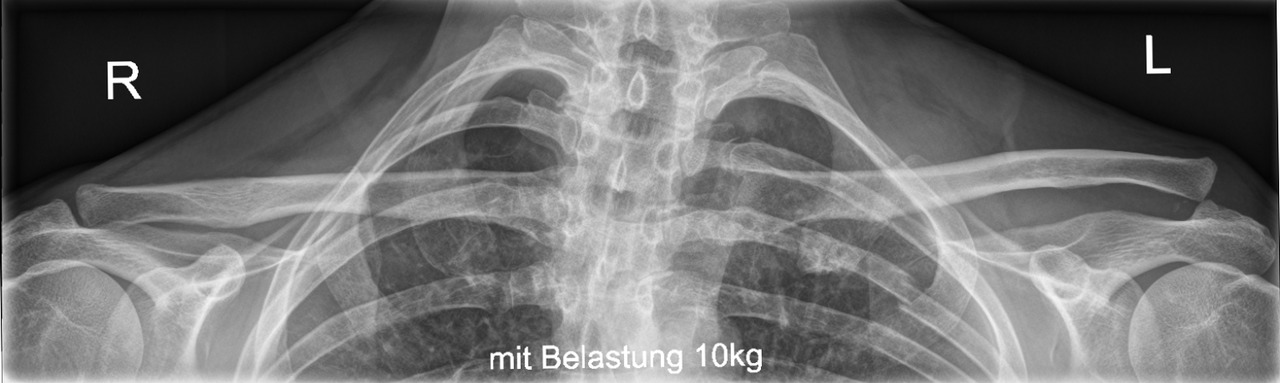
Mechanism
Direct fall onto the tip of the shoulder with the arm adducted (cycling, contact sports).
Clinical findings
- Step at the AC joint, tenderness; piano-key sign (the clavicle can be pushed down and springs back).
- Type IV: posterior displacement felt from above; check the skin over the clavicle in type V.
Imaging
- Zanca view (AP tilted 10–15° up) and axillary view; compare with the other side and measure the coracoclavicular distance.
Classification
- I
- AC sprain, no displacement.
- II
- AC torn, CC intact; less than 25% displacement.
- III
- AC and CC torn; 25–100%.
- IV
- Clavicle displaced posteriorly into the trapezius.
- V
- 100–300% upward displacement, deltotrapezial fascia stripped.
- VI
- Clavicle below the coracoid.
Types shown in 3D
- Rockwood I
- Sprain of the acromioclavicular ligaments; the joint is not displaced.
- Rockwood II
- AC ligaments torn, coracoclavicular (CC) ligaments intact: the clavicle rides up by less than its own thickness.
- Rockwood III
- AC and CC ligaments torn: the lateral clavicle stands up by 25–100% (in reality the shoulder droops under the weight of the arm). Most are treated without surgery.
- Rockwood IV
- The lateral clavicle is driven backwards through or into the trapezius. Seen on the axillary view. Operative.
- Rockwood V
- Gross upward displacement (100–300%): the deltotrapezial fascia is stripped off the clavicle. Operative: hook plate, or coracoclavicular reconstruction.
Treatment
- Types I–II
- Sling for comfort, early movement; return to sport in 2–6 weeks.
- Type III
- Non-operative for most; surgery for heavy labourers, overhead athletes or persistent pain and scapular dyskinesia.
- Types IV–VI
- Reduction and stabilisation: hook plate, coracoclavicular suture-button fixation, or ligament reconstruction (anatomical CC reconstruction) for chronic cases.
Aftercare
Hook plates are removed after about 3 months.
Complications
- Persistent deformity (usually cosmetic)
- AC joint arthritis
- Loss of reduction after fixation
- Coracoid or clavicle fracture after tunnel fixation
See it in 3D on the real bones, displace and reduce it, with simulated X-ray and CT — free, in your browser.
Open in 3D →