 OpenDissect
All fractures › Dislocations
Elbow dislocation
Simple / complexPosterior, posterolateral, terrible triad
See it in 3D on the real bones, displace and reduce it, with simulated X-ray and CT — free, in your browser.
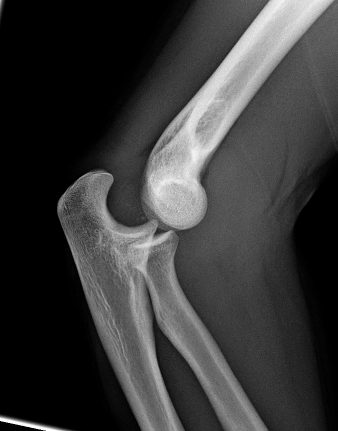
Open in 3D →The second most dislocated large joint in adults. Simple dislocations have no fracture and do well with early movement. Complex dislocations come with fractures (radial head, coronoid, olecranon) and tend to be unstable; the terrible triad is the classic one.
Mechanism
Fall on the outstretched hand: axial load, valgus and supination drive the forearm posterolaterally off the humerus.
Clinical findings
- Deformity with a prominent olecranon, elbow held flexed.
- Check the brachial artery (radial pulse) and the ulnar, median and radial nerves before and after reduction.
- Look at the wrist (DRUJ, Essex–Lopresti) and the shoulder.
Imaging
- AP and lateral before and after reduction: concentric joint? CT for any fracture (radial head, coronoid).
Classification
- Simple
- No fracture (ligaments only).
- Complex
- With fractures: terrible triad, transolecranon, varus posteromedial rotational instability (anteromedial coronoid).
- Direction
- Posterior or posterolateral (most), anterior, medial, lateral, divergent.
- Coronoid (Regan–Morrey)
- I tip, II up to 50%, III more than 50% of the coronoid height.
Types shown in 3D
- Simple posterior
- The most common elbow dislocation (adults: second only to the shoulder). Ulna and radius go back together; no fracture. Reduce, test stability in extension, move early.
- Posterolateral
- The forearm rotates off the humerus around the intact medial collateral ligament (Horii circle: lateral ligament fails first, then the capsule, then the medial side).
- Terrible triad
- Posterior dislocation plus fractures of the radial head and the coronoid process. Very unstable: fix or replace the radial head, fix the coronoid (or its capsule), repair the lateral ligament; medial repair or hinged fixator if still unstable.
Treatment
- Simple
- Closed reduction (traction, correct medial/lateral shift, flex), test stability in extension; short splint then early active movement within the stable arc. Hinged brace if unstable in the last 30°.
- Complex
- Restore the bony stabilisers in order: coronoid (or its capsule), radial head (fixation or replacement, never excision alone in a dislocation), then the lateral ulnar collateral ligament; medial repair or hinged external fixator if still unstable.
Aftercare
Early motion is the key; avoid long immobilisation (stiffness). Heterotopic ossification prophylaxis can be considered after complex injuries.
Complications
- Stiffness (loss of extension)
- Recurrent instability
- Heterotopic ossification
- Ulnar nerve symptoms
- Brachial artery injury (rare)
See it in 3D on the real bones, displace and reduce it, with simulated X-ray and CT — free, in your browser.
Open in 3D →