 OpenDissect
All fractures › Ankle & foot
Talus fractures
AO/OTA 81Neck (Hawkins), body and processes
See it in 3D on the real bones, displace and reduce it, with simulated X-ray and CT — free, in your browser.
Open in 3D →Three fifths of the talus is covered by cartilage and no tendon inserts on it, so its blood supply enters through small areas: the neck (sinus and canal of the tarsus) and the deltoid branch medially. A displaced neck fracture tears these vessels and puts the body at risk of avascular necrosis; the more joints that are dislocated, the higher the risk.
Mechanism
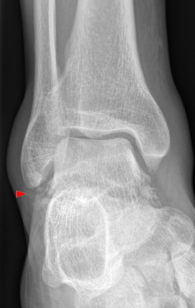
Neck: forced dorsiflexion, the neck levered against the anterior tibial plafond (car footwell, fall from height — the classic "aviator's astragalus"). Body: axial load between plafond and calcaneus. Lateral process: axial load on a dorsiflexed foot with eversion or external rotation (snowboarding). Posterior process: forced plantarflexion or avulsion by the posterior talofibular ligament.
Clinical findings
- Swollen, painful hindfoot; often high-energy with other injuries (calcaneus, spine, pilon).
- Displaced neck and body fractures can tent or perforate the skin, or press on the posterior tibial neurovascular bundle (Hawkins III with posteromedial extrusion): urgent reduction.
- Lateral process fractures mimic an ankle sprain: tenderness just in front of and below the tip of the fibula.
- Posterior process: pain on forced plantarflexion and on moving the big toe (flexor hallucis longus runs between the tubercles).
Imaging
- AP, mortise and lateral of the ankle; AP and oblique of the foot.
- Canale view for the neck (foot in equinus and 15° pronation, tube 15° from vertical).
- CT always: displacement, comminution, process fractures.
- At 6–8 weeks: AP/mortise for the Hawkins sign; MRI if avascular necrosis is suspected.
- AP, mortise and lateral of the ankle plus AP and oblique of the foot.
- Canale view (foot in equinus and 15° pronation, tube tilted 15° from vertical) shows the neck in profile: varus malalignment.
- CT for every suspected talus fracture: displacement, comminution, subtalar involvement and the small process fractures that plain films miss.
- Hawkins sign at 6–8 weeks: a subchondral lucent band under the dome on the AP/mortise view means the body is revascularised (good sign); its absence does not prove necrosis.
Classification
- 81
- Talus: body (including the lateral and posterior processes), neck and head are coded as separate segments, each A/B/C.
- 81.2
- Neck, graded by Hawkins.
- Head
- Rare impaction or shear fractures at the talonavicular joint.
- Hawkins I
- Undisplaced neck fracture. AVN risk under about 10%.
- Hawkins II
- Displaced, subtalar joint subluxed or dislocated, ankle reduced. AVN roughly 10–40% (near zero if the subtalar joint is only subluxed, Vallier IIA; about 25% if dislocated, IIB).
- Hawkins III
- Body dislocated from both ankle and subtalar joints. AVN often above 50%.
- Hawkins IV (Canale–Kelly)
- III plus talonavicular subluxation or dislocation. AVN in most cases.
- Hawkins sign
- Subchondral osteopenia of the dome at 6–8 weeks: evidence of revascularisation.
Types shown in 3D
- Hawkins I
- Undisplaced neck fracture: the ankle and subtalar joints are congruent. Confirm on CT. Cast without weight bearing, or percutaneous screws for early motion.
- Hawkins II
- Displaced neck fracture with the subtalar joint subluxed or dislocated: the foot and the head drift away from the body, which stays in the ankle mortise. Reduce urgently, then ORIF.
- Hawkins III
- The body is dislocated out of both the ankle and the subtalar joints, usually posteromedially behind the medial malleolus, where it threatens the skin and the posterior tibial neurovascular bundle. Emergency open reduction.
- Hawkins IV
- As type III, with the head also subluxed or dislocated at the talonavicular joint (Canale–Kelly). Both fragments are without blood supply.
- Lateral process
- "Snowboarder's fracture": the inferolateral process that carries the lateral subtalar facet breaks off. Easily missed as a sprain; CT shows the size. Large displaced fragments are fixed, small comminuted ones excised.
- Posterior process
- The posterior process (lateral tubercle, sometimes the medial one) breaks off with forced plantarflexion; flexor hallucis longus runs in the groove between the tubercles. Not to be confused with an os trigonum (smooth, rounded edges).
- Body (shear)
- Fracture behind the neck that enters the ankle and subtalar joints, here a sagittal shear of the medial body. Articular step and AVN risk as with neck fractures: ORIF, often through a malleolar osteotomy.
Treatment
- Undisplaced neck or body (Hawkins I)
- Confirm on CT that it is truly undisplaced. Non-weight-bearing cast for 6–8 weeks; some fix percutaneously (posterolateral screws) to allow early motion.
- Displaced neck (Hawkins II–IV)
- Urgent closed reduction of any dislocation to relieve the skin and the neurovascular bundle; definitive anatomical ORIF when soft tissues allow, through combined anteromedial and anterolateral approaches. Medial comminution makes the neck collapse into varus: bridge it with a plate or positional screw, do not compress it. Hawkins III–IV are emergencies.
- Body
- Displaced articular fractures: ORIF, often with a medial malleolar osteotomy for access. Severe comminution: consider primary or later tibiotalocalcaneal fusion.
- Lateral and posterior process
- Small, undisplaced: below-knee cast or boot 4–6 weeks. Large displaced fragments (> 1 cm, > 2 mm step): screw fixation. Small comminuted fragments: excision. Persistent pain from a non-union: late excision.
Aftercare
Non-weight-bearing for 8–12 weeks with early ankle and subtalar motion once wounds heal; watch for the Hawkins sign and follow with X-rays (MRI if AVN is suspected).
Complications
- Avascular necrosis of the body (Hawkins II–IV)
- Subtalar and ankle post-traumatic arthritis (the most common long-term problem)
- Varus malunion of the neck: stiff, supinated foot
- Skin necrosis and infection over displaced fragments
- Missed lateral process fracture: chronic lateral pain
See it in 3D on the real bones, displace and reduce it, with simulated X-ray and CT — free, in your browser.
Open in 3D →