 OpenDissect
All fractures › Ankle & foot
Ankle fracture, Weber C
AO/OTA 44CSuprasyndesmotic, syndesmosis torn
See it in 3D on the real bones, displace and reduce it, with simulated X-ray and CT — free, in your browser.
Open in 3D →The fibula breaks above the syndesmosis. The force has torn the tibiofibular ligaments and usually the deltoid ligament, so the distal fibula and talus drift laterally: the mortise is unstable by definition.
Mechanism
Pronation–external rotation (Lauge-Hansen PER). The extreme form is the Maisonneuve injury: fracture of the proximal fibula with a complete syndesmotic tear.
Clinical findings
- Medial tenderness and swelling; palpate the whole fibula up to the knee (Maisonneuve).
Imaging
- AP, mortise and lateral of the ankle.
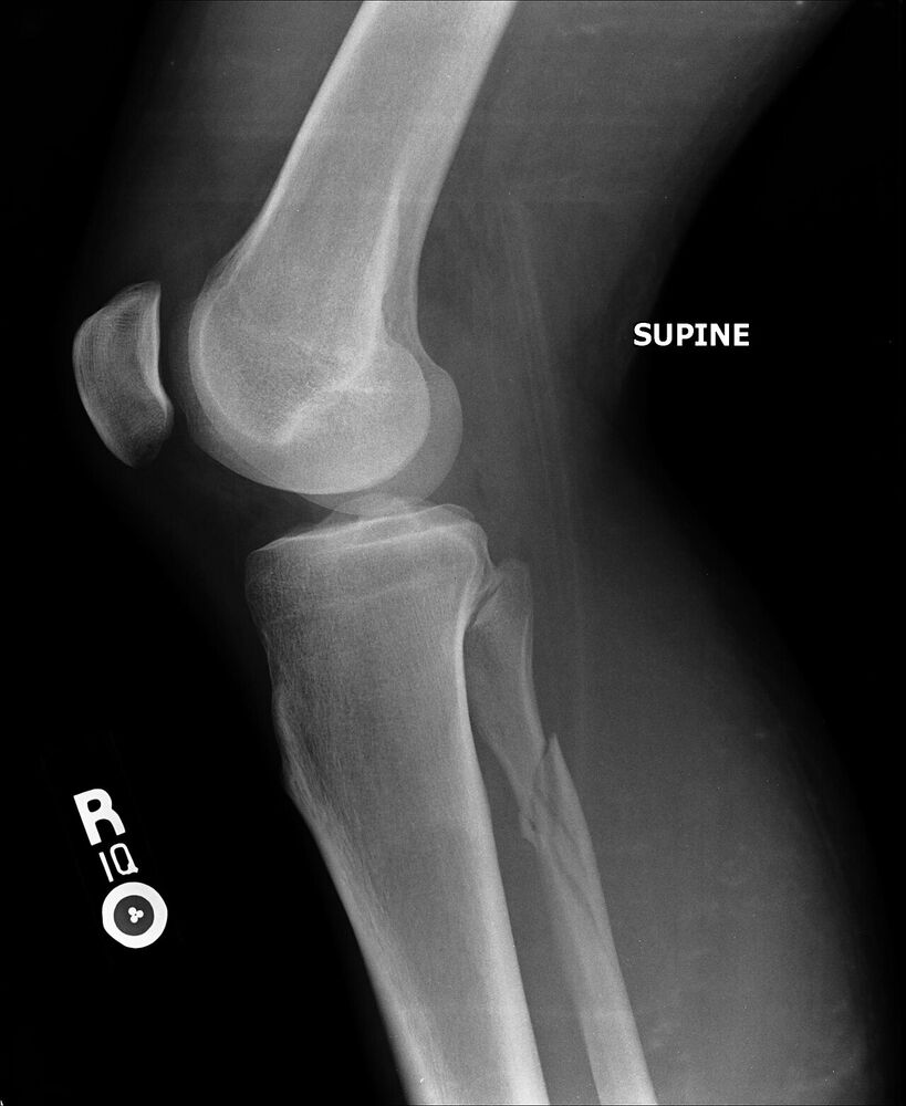
- Full-length tibia and fibula: a proximal fibular fracture (Maisonneuve) means the syndesmosis is torn.
- CT for posterior malleolus and syndesmosis when in doubt.
- Ankle series plus full-length tibia and fibula. Widened tibiofibular clear space (> 6 mm) and medial clear space.
- CT (bilateral) is useful to judge syndesmotic reduction.
Classification
- 44C1
- Simple diaphyseal fibula fracture.
- 44C2
- Multifragmentary fibula.
- 44C3
- Proximal fibula (Maisonneuve).
Types shown in 3D
- 44C1
- Simple diaphyseal fibula fracture above the syndesmosis; syndesmosis and deltoid torn, the talus drifts laterally.
- 44C2
- Multifragmentary fibula above the syndesmosis.
- 44C3
- Proximal fibula (Maisonneuve): the whole interosseous membrane is torn up to the fracture. Fix the syndesmosis; the fibula itself usually not.
Treatment
- Operative (standard)
- Restore fibular length and rotation (plate), then reduce the syndesmosis under direct vision with the ankle in neutral and fix it with one or two 3.5 mm screws or a suture-button device. Repair or fix the medial side if it blocks reduction.
- Maisonneuve
- The fibula itself usually does not need fixation: reduce and fix the syndesmosis.
Aftercare
Non- or partial weight bearing for about 6 weeks; syndesmotic screws are sometimes removed after 8–12 weeks.
Complications
- Syndesmotic malreduction (the main cause of poor results)
- Screw breakage
- Post-traumatic arthritis
See it in 3D on the real bones, displace and reduce it, with simulated X-ray and CT — free, in your browser.
Open in 3D →