 OpenDissect
All fractures › Ankle & foot
Calcaneus fracture
AO/OTA 82Intra-articular, joint depression
See it in 3D on the real bones, displace and reduce it, with simulated X-ray and CT — free, in your browser.
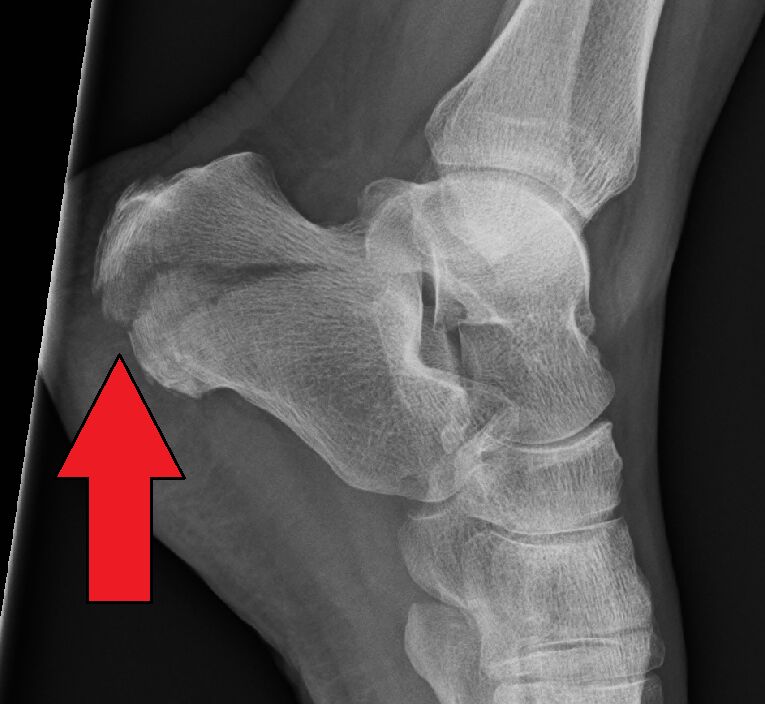
Open in 3D →The talus is driven into the calcaneus like a wedge, breaking the posterior facet of the subtalar joint. The heel widens and shortens; Böhler's angle flattens.
Mechanism
Fall from height onto the heels (look for spine and other lower-limb fractures).
Clinical findings
- Heel swelling and bruising extending to the sole; blisters; compartment syndrome of the foot.
Imaging
- Lateral of the hindfoot: Böhler’s angle (normal 20–40°) and Gissane’s angle.
- Axial (Harris) view: width, varus and the sustentaculum.
- AP of the foot: calcaneocuboid joint.
- CT (axial, coronal and sagittal): mandatory for intra-articular fractures (Sanders classification). Check the spine: same mechanism.
- Lateral (Böhler angle normally 20–40°), axial (Harris) and oblique views; CT is mandatory for intra-articular fractures (Sanders classification on coronal cuts).
Classification
- Extra-articular
- Tuberosity avulsions, anterior process, body without subtalar involvement.
- Sanders I–IV
- Number of posterior facet fragments on coronal CT (I undisplaced, II two parts, III three, IV comminuted).
Types shown in 3D
- Tuberosity avulsion
- Extra-articular: the Achilles tendon pulls off the posterosuperior tuberosity. Emergency if the skin is tented: reduce and fix.
- Tongue type
- Intra-articular (Essex-Lopresti): the secondary line exits through the back of the tuberosity, so the posterior facet stays attached to a "tongue" that tilts up. Can be reduced percutaneously.
- Joint depression (Sanders II–III)
- The posterior facet is a separate fragment driven down into the body: Böhler's angle flattens. Open or sinus-tarsi reduction.
- Sanders IV
- Comminuted posterior facet (four or more parts). Poor results; consider primary subtalar fusion.
Treatment
- Non-operative
- Undisplaced or in smokers, diabetics and vascular disease: elevation, early motion, non-weight-bearing 8–10 weeks.
- Operative
- Displaced intra-articular fractures in suitable patients: sinus tarsi (minimally invasive) or extended lateral approach, restore the facet, height and width. Tuberosity avulsions are emergencies (skin pressure). Sanders IV: consider primary subtalar fusion.
Aftercare
No weight bearing for 8–12 weeks.
Complications
- Wound breakdown (extended lateral approach)
- Subtalar arthritis
- Heel widening, peroneal impingement
- Compartment syndrome → claw toes
See it in 3D on the real bones, displace and reduce it, with simulated X-ray and CT — free, in your browser.
Open in 3D →Related
Ankle fracture, Weber AAO/OTA 44A1Ankle fracture, Weber BAO/OTA 44BAnkle fracture, Weber CAO/OTA 44CFifth metatarsal base fracturesAO/OTA 87.5Talus fracturesAO/OTA 81Navicular fracturesAO/OTA 83Lisfranc injuries and midfoot fracturesAO/OTA 84 cuboid, 85 cuneiforms, 87 metatarsal basesMetatarsal shaft and neck fracturesAO/OTA 87