 OpenDissect
All fractures › Ankle & foot
Fifth metatarsal base fractures
AO/OTA 87.5Avulsion vs Jones fracture
See it in 3D on the real bones, displace and reduce it, with simulated X-ray and CT — free, in your browser.
Open in 3D →Three zones at the base of the fifth metatarsal behave differently: the tuberosity (zone 1) heals reliably, the metaphyseal–diaphyseal junction (zone 2, the true Jones fracture) lies in a watershed area and heals poorly, and the proximal shaft (zone 3) is usually a stress fracture.
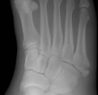
Mechanism
Zone 1: inversion (peroneus brevis and lateral band of the plantar fascia avulse the tip). Zone 2: adduction of the forefoot with the ankle plantarflexed. Zone 3: repetitive load (athletes).
Clinical findings
- Tenderness over the base of the fifth metatarsal (part of the Ottawa foot rules).
Imaging
- AP, oblique and lateral of the foot (Ottawa foot rules).
- Zone matters: avulsion (zone 1), Jones (zone 2) or diaphyseal stress fracture (zone 3).
- AP, oblique and lateral foot. Do not confuse with the apophysis in children (parallel to the shaft) or the os peroneum.
Classification
- Zone 1
- Tuberosity avulsion.
- Zone 2
- Jones fracture at the metaphyseal–diaphyseal junction, extends into the 4th–5th intermetatarsal joint.
- Zone 3
- Proximal diaphyseal stress fracture.
Types shown in 3D
- Zone 1
- Tuberosity avulsion (pseudo-Jones): heals reliably. Stiff shoe, weight bearing as tolerated.
- Zone 2
- Jones fracture at the metaphyseal–diaphyseal junction (into the 4th–5th intermetatarsal joint): watershed blood supply, slow healing. Non-weight-bearing cast or screw.
- Zone 3
- Proximal diaphyseal stress fracture (athletes): screw fixation is often preferred.
Treatment
- Zone 1
- Symptomatic: stiff-soled shoe or boot, weight bearing as tolerated.
- Zone 2–3
- Non-weight-bearing cast for 6–8 weeks, or intramedullary screw fixation in athletes and in delayed union.
Aftercare
Gradual return to sport guided by symptoms and healing.
Complications
- Non-union and refracture (zones 2–3)
See it in 3D on the real bones, displace and reduce it, with simulated X-ray and CT — free, in your browser.
Open in 3D →