 OpenDissect
All fractures › Ankle & foot
Lisfranc injuries and midfoot fractures
AO/OTA 84 cuboid, 85 cuneiforms, 87 metatarsal basesTarsometatarsal fracture-dislocation
See it in 3D on the real bones, displace and reduce it, with simulated X-ray and CT — free, in your browser.
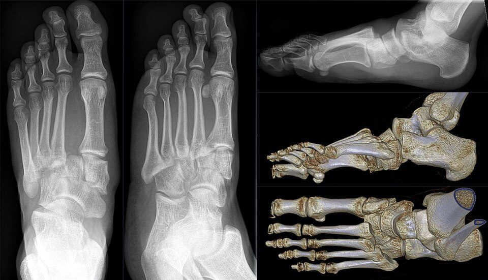
Open in 3D →The base of the second metatarsal is recessed between the medial and lateral cuneiforms like a keystone, and there is no ligament between the first and second metatarsal bases. The Lisfranc ligament (from the medial cuneiform to the plantar base of the second metatarsal) holds the whole forefoot to the midfoot. Injuries range from subtle ligament sprains to complete fracture-dislocations, and about one in five is missed at first presentation.
Mechanism
Indirect: axial load on a plantarflexed foot (missing a step, a rider's foot caught in the stirrup, a player landed on). Direct: crush (heavy object, car). Forced abduction of the forefoot can crush the cuboid between the fourth and fifth metatarsal bases and the calcaneus ("nutcracker").
Clinical findings
- Midfoot pain, swelling, inability to bear weight; plantar ecchymosis over the midfoot is highly suggestive.
- Pain on passive pronation–abduction of the forefoot and on the "piano key" test of the second metatarsal.
- High-energy injuries: check for compartment syndrome of the foot and dorsalis pedis injury (the artery dives between the first and second metatarsal bases).
Imaging
- AP, 30° oblique and lateral of the foot, weight-bearing (or comparison AP of both feet).
- Check the alignment lines of the second and fourth metatarsals and the first–second intermetatarsal space; look for the fleck sign.
- CT for fracture-dislocations; MRI for suspected ligamentous injury with normal films.
- AP, 30° oblique and lateral of the foot, weight-bearing if at all possible (or a comparison AP of both feet).
- AP: medial border of the second metatarsal in line with the medial border of the intermediate cuneiform; diastasis between the first and second metatarsal bases (> 2 mm, or > 1 mm compared with the other side).
- Oblique: medial border of the fourth metatarsal in line with the medial border of the cuboid. Lateral: dorsal step at the tarsometatarsal joints.
- Fleck sign: a small bony fragment in the first intermetatarsal space = Lisfranc ligament avulsion.
- CT for any fracture-dislocation (fragments, cuboid and cuneiform fractures); MRI for purely ligamentous injuries with normal films.
Classification
- Lisfranc
- No separate AO/OTA code: coded by the bones broken (85 cuneiforms, 87 metatarsal bases) plus the dislocation.
- 84
- Cuboid: A extra-articular (avulsion), B partial articular, C complete articular (nutcracker crush).
- 85
- Cuneiforms (medial, intermediate, lateral): A, B, C as for the cuboid.
- Myerson A
- Total incongruity: all five metatarsals displaced in one direction (homolateral), usually lateral.
- Myerson B1 / B2
- Partial incongruity: B1 first ray alone displaced medially; B2 one or more lateral rays displaced laterally.
- Myerson C1 / C2
- Divergent: the first metatarsal goes medially and the others laterally; C1 partial, C2 total.
- Nunley–Vertullo
- Low-energy (athletic) sprains: I no diastasis, II 1–5 mm diastasis compared with the other foot without arch collapse, III diastasis with loss of arch height.
Types shown in 3D
- Fleck sign (subtle)
- Low-energy injury: the Lisfranc ligament pulls a fleck of bone off the medial cuneiform (or the second metatarsal base) and the second metatarsal drifts laterally a few millimetres. Weight-bearing films show the diastasis; if present, it is unstable.
- Myerson A
- Total incongruity: all five metatarsals displaced together, usually laterally and dorsally (homolateral). High energy; check the dorsalis pedis and the compartments.
- Myerson B2
- Partial incongruity: the lateral rays (here second to fifth) are displaced laterally while the first ray stays reduced.
- Myerson C
- Divergent: the first metatarsal goes medially and the others laterally, splitting the first intermetatarsal space (C1 partial, C2 total). Often with intercuneiform and navicular injury.
- Cuboid nutcracker (84C)
- Forced abduction crushes the cuboid between the bases of the fourth and fifth metatarsals and the calcaneus: the lateral column shortens and the forefoot swings into abduction. Restore column length (bridge plate or external fixator, bone graft).
Treatment
- Stable sprain
- No diastasis on weight-bearing films (Nunley I): boot or cast with protected weight bearing for 6 weeks, then a stiff insole.
- Unstable injury
- Any diastasis or malalignment needs anatomical reduction and stable fixation; the result depends on the quality of the reduction. Medial and middle columns (first to third TMT): transarticular screws or dorsal bridge plates (which spare the joint cartilage); the mobile lateral column (fourth and fifth): temporary K-wires. Home-run screw from the medial cuneiform to the second metatarsal base. Purely ligamentous injuries: primary partial arthrodesis of the medial and middle columns is a good alternative (similar or better function and fewer secondary operations, mainly implant removal, in small randomised trials).
- Cuboid and cuneiforms
- Nutcracker cuboid: restore lateral column length with a bridge plate, buttress or external fixator and bone graft for the impaction. Isolated undisplaced cuneiform fractures: cast; displaced or with TMT instability: screws or plates.
- Severe soft tissue
- Compartment release if needed; temporary spanning external fixation and K-wires, definitive fixation when the swelling allows (often 10–14 days).
Aftercare
Non-weight-bearing 6–8 weeks, then progressive loading in a boot; bridge plates and screws are often removed after 4–6 months (not after arthrodesis).
Complications
- Post-traumatic midfoot arthritis (common, worse after a poor reduction)
- Loss of reduction, planovalgus collapse
- Missed diagnosis
- Compartment syndrome
- Dorsalis pedis or deep peroneal nerve injury
See it in 3D on the real bones, displace and reduce it, with simulated X-ray and CT — free, in your browser.
Open in 3D →