 OpenDissect
All fractures › Ankle & foot
Ankle fracture, Weber B
AO/OTA 44BTranssyndesmotic, bimalleolar variant
See it in 3D on the real bones, displace and reduce it, with simulated X-ray and CT — free, in your browser.
Open in 3D →The most common ankle fracture: a short oblique fracture of the fibula starting at the level of the joint and running from anterior-inferior to posterior-superior. Stability depends on the medial side: if the deltoid ligament or the medial malleolus is broken, the talus shifts out of the mortise.
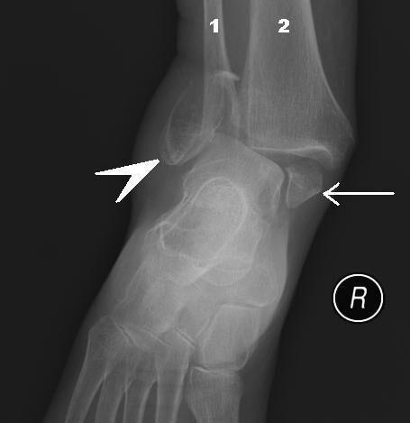
Mechanism
Supination–external rotation (Lauge-Hansen SER): the foot is fixed and the leg rotates internally.
Clinical findings
- Swelling over both malleoli. Medial tenderness, bruising or a medial malleolar fracture suggests instability.
- Check the skin: fracture-dislocations must be reduced immediately to save the skin.
Imaging
- AP, mortise and lateral of the ankle.
- If the medial side looks intact: weight-bearing (or gravity stress) mortise view. Medial clear space ≤ 4 mm and equal to the superior clear space = stable, treat in a boot.
- CT if a posterior malleolar fragment is seen.
- AP, mortise and lateral views. Measure the medial clear space (> 4–5 mm = deltoid injury). If unclear, a weight-bearing or gravity stress view decides.
- Lateral view: look for a posterior malleolus fragment; CT if it looks large.
Classification
- 44B1
- Isolated lateral malleolus (B1.1) or with deltoid rupture.
- 44B2
- With a medial malleolus fracture (bimalleolar).
- 44B3
- With medial and posterior malleolus (trimalleolar).
- Lauge-Hansen SER I–IV
- I anterior syndesmosis, II fibula, III posterior malleolus/posterior syndesmosis, IV medial malleolus or deltoid.
Types shown in 3D
- 44B1
- Isolated transsyndesmotic fibula fracture, medial side intact: the mortise is stable. Walking boot; stress view if in doubt.
- 44B2
- With a medial lesion: medial malleolus fracture (bimalleolar) or deltoid rupture. The talus follows the fibula out of the mortise: fix.
- 44B3
- With a medial lesion and a posterolateral fracture of the tibia (posterior malleolus, Volkmann): trimalleolar. The talus can sublux posteriorly; fix large posterior fragments.
Treatment
- Stable (isolated, stress-negative)
- Walking boot, weight bearing as tolerated for 6 weeks. Repeat X-ray at 1–2 weeks to confirm the mortise.
- Unstable (bi-/trimalleolar or deltoid-equivalent)
- Open reduction and internal fixation: lag screw plus one-third tubular neutralisation plate (lateral) or posterolateral antiglide plate on the fibula; medial malleolus with two partially threaded cancellous screws or a tension band. Test the syndesmosis intraoperatively (hook/Cotton test) and fix it if unstable. Fix large posterior malleolus fragments.
Aftercare
Protected or early weight bearing depending on bone quality and fixation; diabetic and neuropathic patients need longer protection.
Complications
- Wound problems and infection (diabetes, smokers)
- Malreduction of the fibula (shortening, rotation) or of the syndesmosis
- Post-traumatic arthritis
- Symptomatic hardware
See it in 3D on the real bones, displace and reduce it, with simulated X-ray and CT — free, in your browser.
Open in 3D →