 OpenDissect
All fractures › Knee & leg
Tibial plateau fracture
AO/OTA 41B3Lateral split-depression (Schatzker II)
See it in 3D on the real bones, displace and reduce it, with simulated X-ray and CT — free, in your browser.
Open in 3D →The lateral plateau is split and its articular surface pushed down by the lateral femoral condyle in a valgus load. The joint becomes incongruent and unstable, and the lateral meniscus is often trapped in the gap.
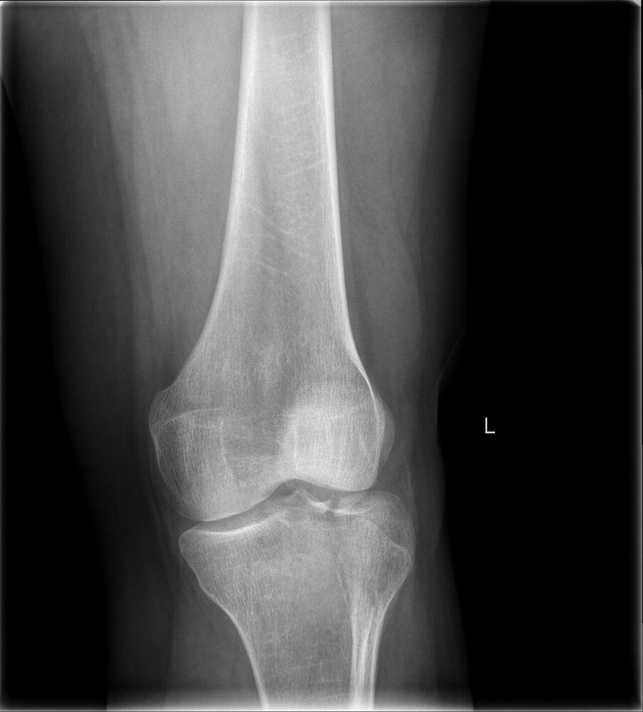
Mechanism
Valgus and axial load ("bumper fracture"); low energy in osteoporotic bone (pure depression), high energy in the young (bicondylar, shaft dissociation).
Clinical findings
- Swollen, painful knee with haemarthrosis (fat droplets on aspiration = lipohaemarthrosis).
- Look for compartment syndrome (especially Schatzker IV–VI) and common fibular nerve injury.
- Test stability in extension under anaesthesia if in doubt.
Imaging
- AP and lateral of the knee.
- Internal and external 45° obliques show the lateral and medial plateaus.
- CT always before surgery: depression, split and fragment map. MRI if a meniscal or ligament injury is suspected.
- Compartment checks and pulses: high-energy patterns (Schatzker IV–VI).
- AP, lateral and oblique X-rays; a cross-table lateral may show a fat–fluid level.
- CT for every plateau fracture to plan the approach; MRI for meniscus and ligaments.
Classification
- Schatzker I
- Lateral split (young, good bone).
- Schatzker II
- Lateral split-depression (most common).
- Schatzker III
- Pure lateral depression (osteoporotic).
- Schatzker IV
- Medial plateau: high energy, often a knee dislocation equivalent: check the popliteal artery.
- Schatzker V
- Bicondylar.
- Schatzker VI
- Plateau dissociated from the shaft.
- AO/OTA 41
- 41A extra-articular, 41B partial articular (B1 split, B2 depression, B3 split-depression), 41C complete articular.
Types shown in 3D
- Schatzker I
- Lateral split, no depression. Young patients with strong bone. Lag screws or buttress plate.
- Schatzker II
- Lateral split-depression (the most common): elevate the depressed joint, graft, and buttress the split.
- Schatzker III
- Pure central depression of the lateral plateau, typical of osteoporotic bone. Elevate through a cortical window and support with graft or raft screws.
- Schatzker IV
- Medial plateau: high energy, a knee-dislocation equivalent. Check the popliteal artery and the common fibular nerve.
- Schatzker V
- Bicondylar: both plateaus split, the shaft still attached in the middle. Staged ex-fix, then dual plating.
- Schatzker VI
- Plateau dissociated from the shaft (metaphyseal–diaphyseal). Highest risk of compartment syndrome.
Treatment
- Non-operative
- Undisplaced, stable fractures: hinged brace, early motion, toe-touch weight bearing for 6–8 weeks.
- Operative
- Articular step > 2–3 mm, condylar widening or instability: elevate the depressed fragment through a cortical window, support it with bone graft or substitute, and buttress the split with a lateral locking plate (or raft screws). Bicondylar fractures: staged ex-fix, then dual plating once the soft tissues allow (wrinkle sign).
Aftercare
Early motion, protected weight bearing 8–12 weeks.
Complications
- Compartment syndrome
- Wound breakdown and infection
- Loss of reduction
- Post-traumatic arthritis
- Stiffness
See it in 3D on the real bones, displace and reduce it, with simulated X-ray and CT — free, in your browser.
Open in 3D →