 OpenDissect
All fractures › Knee & leg
Patella fracture
AO/OTA 34C1Transverse, displaced
See it in 3D on the real bones, displace and reduce it, with simulated X-ray and CT — free, in your browser.
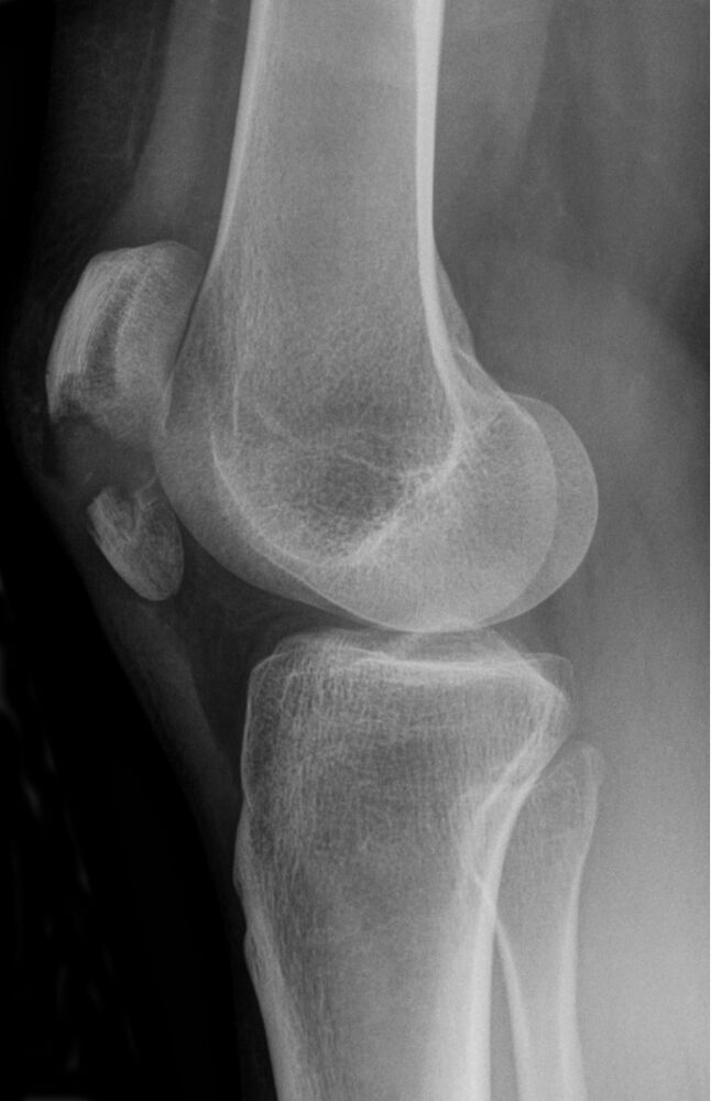
Open in 3D →The patella is the fulcrum of the extensor mechanism. In a transverse fracture the quadriceps pulls the proximal fragment upwards, so the patient cannot actively extend the knee if the retinacula are torn.
Mechanism
Direct blow (dashboard, fall on the knee: comminuted) or sudden eccentric contraction of the quadriceps (transverse).
Clinical findings
- Swollen knee with a palpable gap.
- Straight-leg raise: if the patient cannot lift the extended leg, the extensor mechanism is disrupted.
Imaging
- AP and lateral of the knee (the lateral shows the gap and the step).
- Skyline (axial) view if possible: vertical fractures and the patellofemoral joint.
- CT for comminuted patterns.
- AP, lateral (best for displacement) and skyline views. Do not confuse with a bipartite patella (superolateral, rounded edges, often bilateral).
Classification
- 34A
- Extra-articular (pole avulsion).
- 34B
- Partial articular, vertical.
- 34C
- Complete articular: C1 transverse, C2 transverse with a second fragment, C3 complex.
Types shown in 3D
- 34A
- Extra-articular: avulsion of the inferior pole. Small pole fragments can be excised and the tendon reattached.
- 34B1
- Partial articular, vertical (lateral facet). The extensor mechanism usually works: often non-operative.
- 34C1
- Complete articular, transverse: the quadriceps pulls the proximal fragment up. Tension-band wiring.
- 34C3
- Complete articular, multifragmentary (stellate). Plates, cerclage or partial patellectomy.
Treatment
- Non-operative
- Extensor mechanism intact and displacement < 2–3 mm step or gap: brace in extension, full weight bearing, progressive flexion.
- Operative
- Tension-band wiring (two K-wires or cannulated screws with a figure-of-eight wire) converts the pull of the quadriceps into compression on the articular side. Comminuted patterns: plates or partial patellectomy with reattachment of the tendon. Repair the retinacula.
Aftercare
Early range of motion in a hinged brace; avoid full active extension against resistance for 6 weeks.
Complications
- Symptomatic hardware (very common)
- Loss of reduction
- Patellofemoral arthritis
- Extension lag
See it in 3D on the real bones, displace and reduce it, with simulated X-ray and CT — free, in your browser.
Open in 3D →