 OpenDissect
All fractures › Knee & leg
Tibial pilon fracture
AO/OTA 43CDistal tibial articular (plafond)
See it in 3D on the real bones, displace and reduce it, with simulated X-ray and CT — free, in your browser.
Open in 3D →An axial load drives the talus into the tibial plafond and bursts the articular surface. The soft-tissue envelope is thin and often badly damaged, which dictates the timing of surgery.
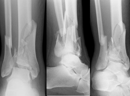
Mechanism
Falls from height and road accidents (axial load); low-energy rotational variants in the elderly.
Clinical findings
- Severe swelling and fracture blisters; open wounds; check for calcaneal and spinal fractures (same mechanism).
Imaging
- AP, mortise and lateral of the ankle; AP and lateral of the whole tibia.
- CT, usually after spanning external fixation (ligamentotaxis makes the fragments easier to read).
- X-rays, then CT after spanning ex-fix to plan fixation (the three classic fragments: medial, anterolateral Chaput, posterolateral Volkmann).
Classification
- 43A
- Extra-articular distal tibia.
- 43B
- Partial articular.
- 43C
- Complete articular (true pilon).
- Rüedi–Allgöwer
- I undisplaced, II displaced without comminution, III impacted and comminuted.
Types shown in 3D
- 43A
- Extra-articular distal tibia (with fibula). Nail with distal locking or plate.
- 43B1
- Partial articular, pure split (here the medial plafond).
- 43C
- Complete articular: the joint surface breaks into the classic medial, anterolateral (Chaput) and posterolateral (Volkmann) fragments, and the talus is driven up between them.
Treatment
- Staged protocol
- Day 0: spanning external fixator (± fibular fixation) to restore length. When the swelling settles (often 7–21 days, wrinkle sign): anatomical reduction of the joint and plate fixation through approaches planned on CT.
- Alternatives
- Definitive ex-fix with limited internal fixation when the soft tissues are poor; primary arthrodesis in unreconstructable fractures.
Aftercare
Early ankle motion, non-weight-bearing 10–12 weeks.
Complications
- Wound necrosis and deep infection
- Post-traumatic arthritis (very common)
- Malunion, non-union
See it in 3D on the real bones, displace and reduce it, with simulated X-ray and CT — free, in your browser.
Open in 3D →