 OpenDissect
All fractures › Knee & leg
Proximal fibula fracture & Maisonneuve injury
AO/OTA 4F1 · 44C3Common peroneal nerve · syndesmosis
See it in 3D on the real bones, displace and reduce it, with simulated X-ray and CT — free, in your browser.
Open in 3D →An isolated break of the proximal fibula is usually harmless to stability, because the fibula carries little load. Two things make it important: the common peroneal nerve winds around the fibular neck just below the head, and a proximal fibula fracture after an ankle twist may be the only visible part of a Maisonneuve injury, in which the force has torn the deltoid ligament (or broken the medial malleolus), the syndesmosis and the interosseous membrane up to the fracture.
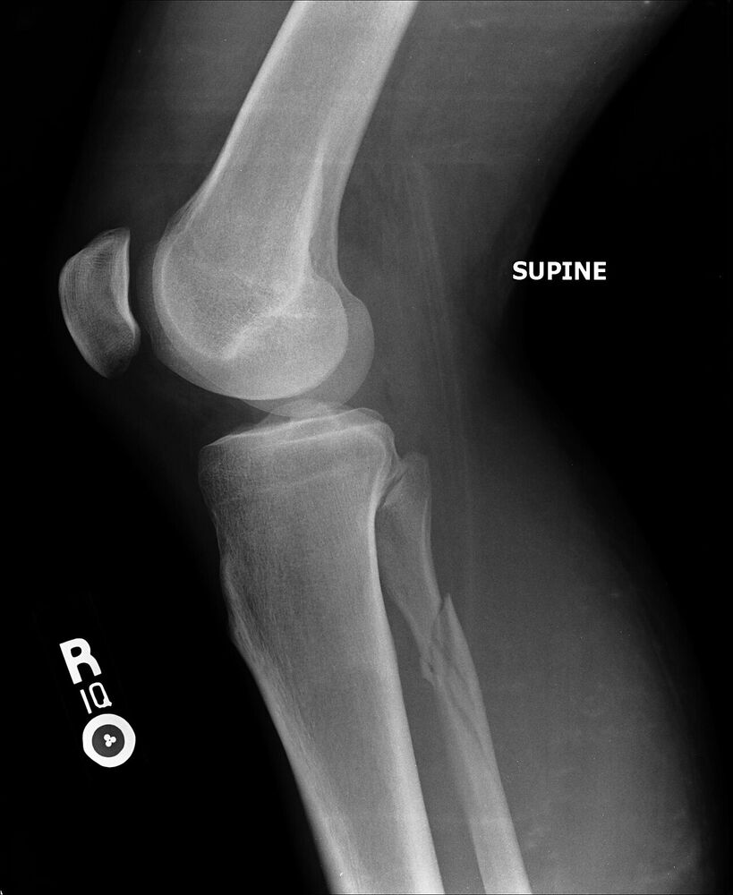
Mechanism
Direct blow to the lateral knee (bumper, kick): head or neck fracture. Varus and hyperextension of the knee: avulsion of the fibular head or styloid by the lateral collateral ligament and biceps femoris (“arcuate” fracture, a marker of posterolateral corner injury). Pronation–external rotation of the ankle: Maisonneuve.
Clinical findings
- Tenderness over the fibular head or neck. Test ankle dorsiflexion, toe extension, eversion and sensation in the first web space (common peroneal nerve).
- After any ankle injury, palpate the whole fibula up to the knee: proximal tenderness with medial ankle tenderness or swelling means Maisonneuve until proven otherwise.
- Fibular head avulsion: examine the knee for posterolateral corner and cruciate injuries (dial test, varus at 30°, posterior drawer).
Imaging
- AP and lateral of the knee for fibular head or neck fractures.
- After any ankle injury with proximal fibular tenderness: AP, mortise and lateral of the ankle plus full-length tibia and fibula.
- Mortise view: medial clear space (> 4–5 mm) and tibiofibular clear space (> 6 mm) mean syndesmotic and deltoid injury; stress or weight-bearing view if borderline. MRI of the knee for an arcuate (styloid) avulsion.
- AP and lateral of the knee for head and neck fractures; AP, mortise and lateral of the ankle plus full-length tibia and fibula whenever the ankle hurts.
- Maisonneuve: widened medial clear space (> 4–5 mm) and tibiofibular clear space (> 6 mm) on the mortise view; if normal, a stress or weight-bearing view. Bilateral CT helps judge syndesmotic reduction.
- Fibular styloid avulsion (arcuate sign): MRI of the knee for the posterolateral corner and cruciates.
Classification
- 4F1A
- Proximal fibula, simple.
- 4F1B
- Proximal fibula, multifragmentary.
- Arcuate fracture
- Avulsion of the fibular styloid (arcuate and popliteofibular ligaments) or lateral head (LCL, biceps femoris): a posterolateral corner injury.
- Maisonneuve (44C3)
- Proximal fibula fracture with complete syndesmotic disruption and a medial injury (deltoid rupture or medial malleolus fracture): an unstable ankle.
Types shown in 3D
- Fibular head avulsion
- The fibular styloid (popliteofibular and arcuate ligaments) or the lateral head (LCL and biceps femoris) is pulled off (arcuate fracture). Small fragment, big meaning: a posterolateral corner injury, often with cruciate tears.
- 4F1A neck
- Simple fracture of the fibular neck from a direct blow to the lateral knee. Barely displaced, because the tibia splints the fibula; check the common peroneal nerve, which crosses the neck here.
- Maisonneuve
- Spiral fracture of the proximal third of the fibula from external rotation of the foot. The syndesmosis and deltoid ligament are torn, so the distal fibula and the talus rotate externally and drift laterally out of the mortise.
- Maisonneuve + medial malleolus
- The medial injury is a malleolar fracture instead of a deltoid tear. Fix the medial malleolus and the syndesmosis; the proximal fibula is left alone.
Treatment
- Isolated head or neck fracture
- Symptomatic: weight bearing as tolerated, brace for comfort. Document the peroneal nerve; a palsy is observed (most are neurapraxias) with an ankle–foot orthosis, and explored if it fails to recover by about 3 months on EMG.
- Fibular head avulsion with posterolateral corner injury
- Early (within about 2–3 weeks) repair of the avulsed fragment with screw, suture anchors or a cerclage, together with reconstruction of the posterolateral corner and cruciates as needed.
- Maisonneuve
- The proximal fibula itself is not fixed (risk to the nerve). Restore fibular length and rotation at the ankle, reduce the syndesmosis under direct vision or with bilateral fluoroscopic comparison, and fix it with one or two screws or suture-button devices. Fix a medial malleolar fracture; the deltoid is repaired if it blocks reduction.
Aftercare
Isolated fractures: full weight bearing as pain allows. Maisonneuve: protected weight bearing for about 6 weeks, then progressive loading; screws are sometimes removed after 8–12 weeks.
Complications
- Common peroneal nerve palsy (foot drop)
- Missed Maisonneuve injury with chronic syndesmotic instability
- Syndesmotic malreduction
- Missed posterolateral corner injury (persistent varus instability)
See it in 3D on the real bones, displace and reduce it, with simulated X-ray and CT — free, in your browser.
Open in 3D →