 OpenDissect
All fractures › Cervical spine
Subaxial cervical injuries (C3–C7)
AO Spine subaxial cervicalCompression, tension band, translation, facet dislocation
See it in 3D on the real bones, displace and reduce it, with simulated X-ray and CT — free, in your browser.
Open in 3D →From C3 to C7 the cervical spine fails in three basic ways: the vertebral body is crushed (A), the posterior or anterior tension band is torn (B), or one vertebra translates on the next (C), typically as a facet dislocation. C5–C7 are most often affected. Every injury is graded together with the neurological status and the facets.
Mechanism
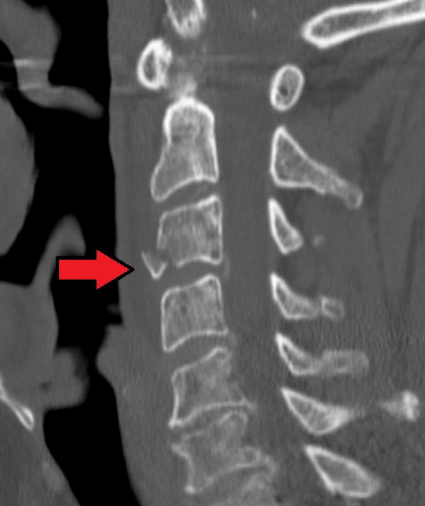
Flexion with axial load (diving, rugby, rollover crashes): compression, burst and teardrop fractures. Flexion–distraction and rotation: facet subluxation and dislocation. Hyperextension: anterior tension band failure, especially in the stiff spine (ankylosing spondylitis, DISH) and in the elderly with spondylosis (central cord syndrome).
Clinical findings
- Spinal motion restriction; assess with the Canadian C-spine rule or NEXUS criteria. ATLS primary survey; neurogenic shock (hypotension with bradycardia) differs from haemorrhagic shock.
- Full ISNCSCI/ASIA examination including sacral sparing (perianal sensation, voluntary anal contraction); priapism suggests cord injury.
- A stiff, ankylosed spine fractures with trivial trauma and is highly unstable: image the whole spine and keep the patient in their usual posture.
Imaging
- CT occiput to T1 (radiographs only if CT is unavailable; the lateral must show the C7–T1 junction).
- MRI for any neurological deficit, suspected disc or ligament injury, or before open reduction in a patient who cannot be examined.
- CT angiography for fractures through a transverse foramen, facet dislocations or posterior circulation symptoms.
- CT from occiput to T1 is the first test in adults; radiographs only if CT is unavailable.
- MRI for any neurological deficit, before open reduction of a dislocation in a patient who cannot be examined, and when ligament or disc injury is suspected.
- Facet dislocation: unilateral gives about 25% anterior translation with rotation, bilateral about 50%. Look for the "naked facet" on axial CT.
- CT angiography for fractures through the transverse foramen and facet dislocations (vertebral artery injury).
Classification
- A0–A4
- Compression injuries: A0 minor (spinous or transverse process), A1 one endplate, A2 coronal split or pincer, A3 burst with one endplate, A4 burst with both endplates or split with posterior wall.
- B1–B3
- Tension band injuries: B1 posterior bony, B2 posterior capsuloligamentous or bony-ligamentous, B3 anterior (hyperextension, through the disc or body).
- C
- Translation of one vertebra on another in any plane, including facet dislocations.
- F1–F4
- Facet modifiers: F1 non-displaced facet fracture, F2 facet fracture with potential instability, F3 floating lateral mass, F4 subluxed, perched or dislocated facet (BL = bilateral).
- N, M
- Neurology N0–N4 (and NX); case modifiers M1 posterior ligament injury without complete disruption, M2 critical disc herniation, M3 stiff or metabolic bone disease, M4 vertebral artery abnormality.
Types shown in 3D
- A1 wedge (C5)
- Compression of the upper endplate with an intact posterior wall and tension band. Stable: collar.
- A4 burst (C5)
- Both endplates and the posterior wall fail; a fragment of the posterior wall is pushed back into the narrow cervical canal. Anterior corpectomy and fusion if the cord is compressed.
- B2 posterior tension band (C5–C6)
- Flexion–distraction tears the facet capsules, ligamentum flavum and interspinous ligaments: the spinous processes fan apart, the facets are subluxed but not dislocated. Posterior fixation.
- C bilateral facet dislocation (C5–C6)
- C5 has translated forwards by about half a vertebral body: both inferior facets of C5 are locked in front of the superior facets of C6. High rate of complete cord injury.
- C unilateral facet dislocation (F4)
- Flexion with rotation: one facet jumps and locks, giving about 25% forward slip with rotation of C5 on C6. Often a root deficit rather than cord injury.
Treatment
- Stable (A0–A2, minor F1)
- Rigid collar 6–12 weeks, upright radiographs to confirm alignment.
- Burst and teardrop with cord compression
- Anterior corpectomy and fusion with plate (posterior fixation added for posterior ligament injury). Early decompression (within 24 h) is advised for acute cord injury; keep mean arterial pressure at least 75–80 mmHg (upper target 90–95) for 3–7 days; many centres still aim for 85–90.
- Facet dislocation (C, F4)
- Awake, cooperative and examinable patient: closed reduction with skull tongs and incremental weights under serial neurological examination. Otherwise, or if closed reduction fails, MRI first; if a herniated disc is found, anterior discectomy before reduction. Fusion afterwards, anterior or posterior.
- Tension band injuries (B)
- Usually surgical: posterior lateral mass screws for B1–B2; long posterior fixation for B3 injuries in an ankylosed spine.
Aftercare
Collar after anterior fusion for comfort; rehabilitation in a spinal cord unit for any cord injury. Watch for dysphagia and hoarseness (recurrent laryngeal nerve) after anterior surgery.
Complications
- Neurological deterioration (missed instability, reduction against a disc herniation)
- Vertebral artery injury and posterior circulation stroke
- Adjacent segment degeneration after fusion
- Complications of cord injury: pneumonia, pressure sores, thromboembolism, autonomic dysreflexia
See it in 3D on the real bones, displace and reduce it, with simulated X-ray and CT — free, in your browser.
Open in 3D →