 OpenDissect
All fractures › Cervical spine
Atlas (C1) fractures
AO Spine upper cervical IIJefferson burst, arch and lateral mass
See it in 3D on the real bones, displace and reduce it, with simulated X-ray and CT — free, in your browser.
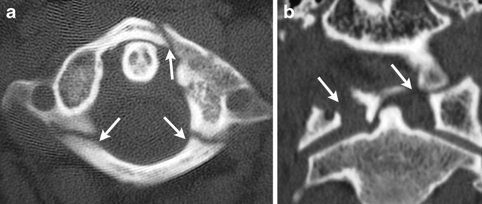
Open in 3D →The atlas is a ring with no body: two lateral masses joined by thin anterior and posterior arches. An axial load drives the occipital condyles down into the lateral masses and bursts the ring outwards. Because the canal widens, the cord is usually spared; the question that decides treatment is whether the transverse atlantal ligament has failed.
Mechanism
Axial load on the vertex: diving into shallow water, falls onto the head, objects falling on the head. Hyperextension crushes the posterior arch between occiput and C2. In the elderly, simple falls.
Clinical findings
- Upper neck pain, often with occipital pain or headache; neurology usually intact.
- Look for a second spinal fracture: about half have another cervical injury, most often of C2 (dens).
- Occipital neuralgia (greater occipital nerve) and vertebral artery injury are possible; check posterior circulation symptoms.
Imaging
- CT of the cervical spine, occiput to T1 (first-line imaging in adults who fail the Canadian C-spine rule or NEXUS).
- Open-mouth view or coronal CT: total lateral mass overhang of C1 on C2 (rule of Spence).
- MRI of the craniocervical junction for the transverse ligament; CT angiography if a transverse foramen is involved.
- CT of the whole cervical spine is the first test; most fractures are only seen clearly on CT.
- Open-mouth view or coronal CT: lateral masses overhanging C2 on both sides add up to the total displacement (rule of Spence).
- MRI shows the transverse ligament directly; CT angiography if the fracture runs into a transverse foramen.
Classification
- Gehweiler 1
- Isolated anterior arch fracture (sometimes a horizontal avulsion by longus colli).
- Gehweiler 2
- Isolated posterior arch fracture, usually bilateral at the vertebral artery groove. Stable.
- Gehweiler 3
- Combined anterior and posterior arch fractures (Jefferson burst): 3a transverse ligament intact (stable), 3b ligament torn (unstable).
- Gehweiler 4
- Lateral mass fracture.
- Gehweiler 5
- Isolated transverse process fracture.
- Dickman
- Transverse ligament injury: type I tear within the ligament (will not heal: fusion), type II bony avulsion of its tubercle on the lateral mass (can heal with immobilisation).
Types shown in 3D
- Gehweiler 2 posterior arch
- Bilateral fracture of the thin posterior arch at the vertebral artery grooves, crushed between occiput and C2 in hyperextension. Stable: collar.
- Gehweiler 3a Jefferson, ligament intact
- Four-part burst of the ring (two anterior, two posterior arch fractures). The lateral masses spread only slightly: the transverse ligament holds. Collar.
- Gehweiler 3b Jefferson, ligament torn
- The lateral masses are driven far apart: a combined overhang on C2 of more than 6.9 mm on CT (rule of Spence) suggests the transverse ligament has failed. MRI decides between avulsion (Dickman II) and tear (Dickman I).
- Gehweiler 4 lateral mass
- The condyle splits one lateral mass; the outer part with the transverse process is pushed down and out, usually with an anterior arch fracture on the same side. Risk of late C0–C1 arthritis and head tilt.
Treatment
- Stable (arch fractures, Jefferson with intact ligament)
- Rigid collar for about 8–12 weeks with upright X-rays or CT to confirm alignment; flexion–extension films after healing.
- Transverse ligament avulsion (Dickman II)
- Immobilisation (rigid collar, or halo in selected young patients) usually heals; check stability at about 3 months and fuse C1–C2 if it does not.
- Unstable (Dickman I, widely displaced burst)
- Posterior C1–C2 fusion (C1 lateral mass and C2 pedicle or pars screws, or transarticular screws). In young patients a direct C1 ring osteosynthesis that keeps C1–C2 motion is an option. Combined C1–C2 fractures are treated according to the C2 injury.
Aftercare
Collar until bony union; avoid contact sports for several months. After C1–C2 fusion about half of neck rotation is lost.
Complications
- Atlantoaxial instability or non-union
- Loss of rotation, chronic neck pain
- Occipital neuralgia
- Vertebral artery injury (rare)
See it in 3D on the real bones, displace and reduce it, with simulated X-ray and CT — free, in your browser.
Open in 3D →