 OpenDissect
All fractures › Cervical spine
Hangman’s fracture (traumatic spondylolisthesis of C2)
AO Spine upper cervical IIILevine–Edwards I, II, IIA, III
See it in 3D on the real bones, displace and reduce it, with simulated X-ray and CT — free, in your browser.
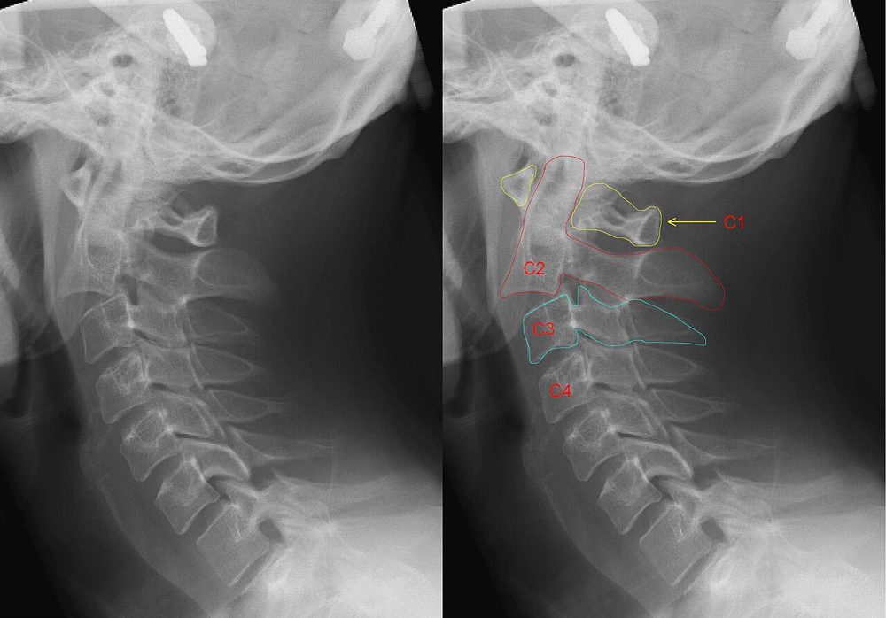
Open in 3D →Bilateral fractures through the pars interarticularis of C2 separate the body of C2 (with the dens, the atlas and the skull) from its posterior arch, which stays attached to C3. The canal widens as the fragments separate, so most patients are neurologically intact. Stability depends on the C2–C3 disc and facets.
Mechanism
Today mostly road traffic accidents and falls: hyperextension with axial load (type I), followed by flexion when the C2–C3 disc tears (type II); flexion–distraction (IIA); flexion–compression with facet dislocation (III). Judicial hanging with a submental knot produced hyperextension with distraction.
Clinical findings
- Neck pain after a head-first impact; facial or forehead injuries suggest hyperextension.
- Neurology usually intact ("self-decompressing" fracture); atypical patterns running through the back of the C2 body are more dangerous for the cord.
- Vertebral artery injury is possible when the line crosses the transverse foramen.
Imaging
- CT of the cervical spine (whole spine in high-energy trauma).
- Lateral view or sagittal CT: translation and angulation of C2 on C3.
- MRI of the C2–C3 disc and ligaments when stability is uncertain; CT angiography if a transverse foramen is involved.
- CT of the cervical spine (whole spine in high-energy trauma).
- The lateral view or sagittal CT shows translation of C2 on C3 (measured at the posterior body margins) and angulation of the C2–C3 disc space.
- MRI to look at the C2–C3 disc and ligaments when deciding between non-operative and operative care; CT angiography if a transverse foramen is involved.
Classification
- Type I
- Translation < 3 mm, no angulation. Stable.
- Type II
- Translation > 3 mm and significant angulation; the C2–C3 disc and posterior longitudinal ligament are torn.
- Type IIA
- Severe angulation with little translation (flexion–distraction); the fracture line is more oblique. Traction makes it worse.
- Type III
- Type I fracture plus bilateral C2–C3 facet dislocation. Always unstable.
Types shown in 3D
- Type I
- Hairline bilateral pars fractures, translation under 3 mm and no angulation. The C2–C3 disc is intact: stable, collar.
- Type II
- The C2 body slides forwards (> 3 mm) and tilts into flexion: the C2–C3 disc and posterior longitudinal ligament are torn. Traction in slight extension and halo, or fixation.
- Type IIA
- Severe angulation with little translation: the disc opens at the back like a hinge (flexion–distraction). Traction is contraindicated; reduce with gentle extension and compression.
- Type III
- Pars fractures plus bilateral C2–C3 facet dislocation: the posterior arch of C2 also jumps forwards over C3. Open reduction and posterior fixation.
Treatment
- Type I
- Rigid collar for about 6–12 weeks.
- Type II
- Reduction (traction with slight extension) and halo for 6–12 weeks, or surgery, increasingly favoured: C2 pars/pedicle screws (direct osteosynthesis), anterior C2–C3 discectomy and fusion, or posterior C1–C3 fixation.
- Type IIA
- No traction: gentle extension and compression under fluoroscopy, then halo, or surgical fixation.
- Type III
- Open reduction of the facet dislocation and posterior fixation (often with C2–C3 fusion); the pars fractures are stabilised at the same time.
Aftercare
Collar or halo until union, with upright films; flexion–extension views at the end of treatment to exclude residual C2–C3 instability.
Complications
- C2–C3 instability or non-union
- Loss of reduction in a halo
- Vertebral artery injury
- Late C2–C3 degeneration and neck pain
See it in 3D on the real bones, displace and reduce it, with simulated X-ray and CT — free, in your browser.
Open in 3D →