 OpenDissect
All fractures › Cervical spine
Odontoid (dens) fractures
AO Spine upper cervical IIIAnderson–D’Alonzo I–III
See it in 3D on the real bones, displace and reduce it, with simulated X-ray and CT — free, in your browser.
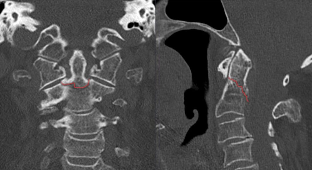
Open in 3D →The most common cervical fracture in people over 70, and a frequent injury in young adults after high-energy trauma. Where the line runs matters: at the narrow waist of the dens (type II) the blood supply is poor and the contact area is small, so non-union is common; through the cancellous body of C2 (type III) it usually heals.
Mechanism
Elderly: a fall from standing onto the face or forehead, often in osteoporotic or spondylotic necks (hyperextension, posterior displacement). Young: high-energy flexion or extension (road traffic, falls).
Clinical findings
- High neck pain; the patient may hold the head in the hands. Neurological deficit is uncommon but can be fatal when the cord is compressed at C1–C2.
- In the elderly the fracture is easily missed: image the neck after any fall onto the head with neck pain or in anyone who cannot be assessed.
- Look for an associated atlas fracture.
Imaging
- CT of the cervical spine with sagittal and coronal reformats.
- Open-mouth and lateral radiographs for follow-up of alignment and healing.
- MRI for neurological signs or to assess the transverse ligament (if an anterior screw is planned).
- CT with sagittal and coronal reformats is the standard. Radiographs (open-mouth and lateral) miss a large share in the elderly.
- Measure displacement and angulation of the dens; posterior displacement and > 5 mm displacement predict non-union.
- MRI for neurological signs or to assess the transverse ligament. Do not confuse with os odontoideum (smooth, corticated, rounded ossicle) or, in young children, the subdental synchondrosis.
Classification
- Type I
- Oblique avulsion of the tip by the alar ligaments. Rare; can be part of an occipitocervical dissociation.
- Type II
- Through the waist, at the junction with the C2 body. The most common and the most prone to non-union.
- Type III
- Extends down into the cancellous body of C2 (in the modified definition, into a superior articular facet).
- Grauer (type II)
- IIA undisplaced transverse; IIB displaced, from anterior-superior to posterior-inferior (suits an anterior screw); IIC from anterior-inferior to posterior-superior or comminuted (posterior fusion).
Types shown in 3D
- Type I (tip)
- Oblique avulsion of the tip above the transverse ligament by the alar ligaments. Stable in itself, but exclude occipitocervical dissociation.
- Type II (waist)
- At the base of the dens, where blood supply and contact area are smallest. Here the dens, the atlas and the head displace backwards together (typical of a fall onto the face in the elderly).
- Grauer IIA (undisplaced)
- Transverse type II fracture without displacement or comminution: suitable for a collar or halo, with close radiological follow-up.
- Type III (into the body)
- The line dips into the cancellous body of C2 and the superior facets. Large, well-vascularised contact area: usually heals in a collar or halo. Here mild anterior displacement in flexion.
Treatment
- Type I and type III
- Rigid collar for about 12 weeks (halo for displaced type III in selected young patients). Rule out occipitocervical instability with type I.
- Type II, young or fit patient
- Surgery if displaced or at high risk of non-union: anterior odontoid screw (fresh fracture, favourable obliquity, intact transverse ligament, good bone and a body habitus that allows the trajectory) or posterior C1–C2 fusion. Undisplaced fractures can be treated in a collar or halo with close follow-up.
- Type II, elderly patient
- Shared decision: a rigid collar often leads to a stable fibrous union that is well tolerated; posterior C1–C2 fusion gives more bony union and may improve survival in fit patients. Halo vests are poorly tolerated in the elderly (pneumonia, falls, swallowing problems) and are avoided.
Aftercare
Upright radiographs or CT at 6 and 12 weeks; flexion–extension views at the end of treatment. Dysphagia is common after anterior screws in the elderly.
Complications
- Non-union (type II: older age, displacement > 5 mm, posterior displacement, angulation, delay)
- Late myelopathy from an unstable non-union
- Dysphagia and aspiration (collars, anterior surgery)
- Vertebral artery injury during C1–C2 screw placement
See it in 3D on the real bones, displace and reduce it, with simulated X-ray and CT — free, in your browser.
Open in 3D →