 OpenDissect
All fractures › Shoulder & arm
Proximal humerus fracture
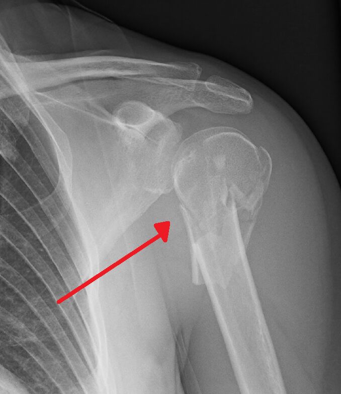
AO/OTA 11BThree-part: surgical neck + greater tuberosity
See it in 3D on the real bones, displace and reduce it, with simulated X-ray and CT — free, in your browser.
Open in 3D →Very common in older women with osteoporosis. The muscles explain the displacement: the supraspinatus and infraspinatus pull the greater tuberosity up and back, the subscapularis pulls the lesser tuberosity medially, and the pectoralis major pulls the shaft medially. The head's blood supply comes mainly from the posterior circumflex humeral artery.
Mechanism
Fall onto the outstretched arm or the side of the shoulder; high-energy injuries and seizures in the young (look for posterior dislocation).
Clinical findings
- Painful, swollen shoulder with extensive bruising after a few days.
- Test the axillary nerve (sensation over the lateral deltoid) and the radial pulse before and after any manipulation.
Imaging
- Trauma series of the shoulder: true AP (Grashey), scapular Y view and axillary view.
- The axillary (or a Velpeau view if the arm cannot be moved) rules out a dislocation.
- CT for 3- and 4-part fractures and head-split patterns before planning.
- Trauma series: true AP (Grashey), scapular Y and axillary views. CT for 3- and 4-part fractures, head-split and fracture-dislocations.
Classification
- Neer
- A "part" is displaced > 1 cm or angulated > 45°. 1-part (undisplaced), 2-part (surgical neck or one tuberosity), 3-part, 4-part, fracture-dislocation, head split.
- AO/OTA 11A
- Extra-articular unifocal (one tuberosity or surgical neck).
- AO/OTA 11B
- Extra-articular bifocal/three-part.
- AO/OTA 11C
- Articular or four-part, anatomical neck.
- Hertel
- Short calcar (< 8 mm) and a disrupted medial hinge (> 2 mm) predict head ischaemia.
Types shown in 3D
- Neer 2-part GT
- Greater tuberosity alone (AO 11A1), pulled up and back by supraspinatus and infraspinatus. More than 5 mm displacement in active patients: fix (screws or sutures).
- Neer 2-part neck
- Surgical neck (AO 11A2–A3): pectoralis major pulls the shaft medially and forwards. Most heal in a sling; plate or nail if displaced in active patients.
- Neer 3-part
- Surgical neck plus greater tuberosity (AO 11B): the head turns into internal rotation because subscapularis is unopposed. Locking plate (PHILOS) with calcar screws and cuff sutures, or reverse prosthesis in the elderly.
- Neer 4-part
- Head, both tuberosities and shaft all separated (AO 11C3). The head often loses its blood supply: reverse shoulder arthroplasty in the elderly.
- Valgus-impacted 4-part
- The head is tilted into valgus and impacted onto the shaft; the medial periosteal hinge is often intact, so the blood supply is better than in classic 4-part fractures. Many can be fixed or treated without surgery.
Treatment
- Non-operative
- Most (about 80%): undisplaced and many displaced fractures in elderly low-demand patients. Sling 2–3 weeks, early pendulum exercises, physiotherapy. Valgus-impacted fractures do well.
- Fixation
- Displaced 2- and 3-part fractures in active patients with reasonable bone: locking plate (PHILOS) through a deltopectoral or deltoid-split approach, restoring the medial calcar (calcar screws) and suturing the cuff to the plate; or an intramedullary nail for surgical neck fractures.
- Replacement
- Unreconstructable 4-part fractures, head-split and fracture-dislocations in the elderly: reverse shoulder arthroplasty (tuberosity repair improves rotation). Hemiarthroplasty in younger patients is now rarely used.
Aftercare
Early passive motion, active from 6 weeks.
Complications
- Screw cut-out into the joint (varus collapse)
- Avascular necrosis
- Axillary nerve injury
- Stiffness
- Tuberosity non-union
See it in 3D on the real bones, displace and reduce it, with simulated X-ray and CT — free, in your browser.
Open in 3D →