 OpenDissect
All fractures › Shoulder & arm
Clavicle fracture
AO/OTA 15.2Midshaft, displaced
See it in 3D on the real bones, displace and reduce it, with simulated X-ray and CT — free, in your browser.
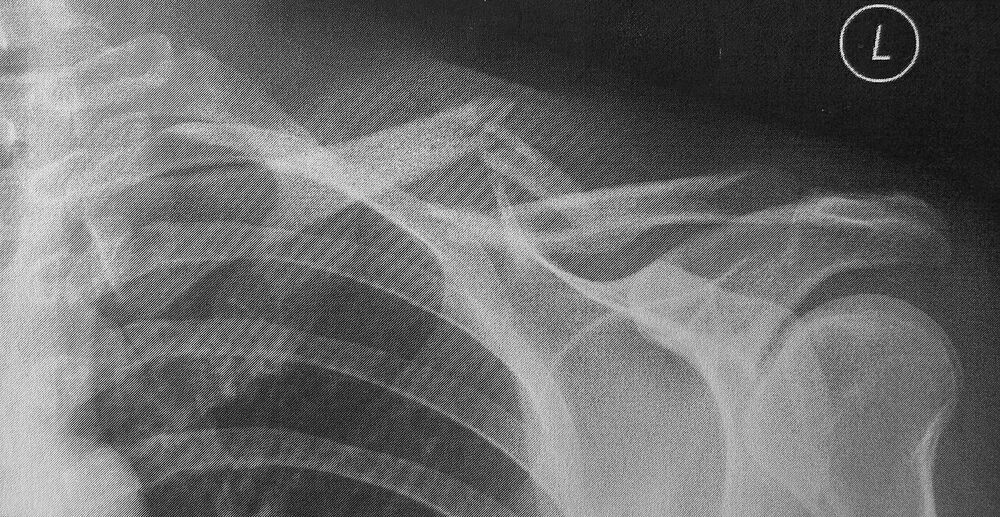
Open in 3D →The most common fracture of the shoulder girdle, usually in the middle third. The sternocleidomastoid lifts the medial fragment; the weight of the arm and pectoralis major pull the lateral fragment down and medially, shortening the clavicle.
Mechanism
Fall onto the shoulder (most), sometimes onto the outstretched hand; sports and cycling.
Clinical findings
- Deformity and tenting of the skin. Check skin integrity, the brachial plexus and the subclavian vessels.
- Look for a "floating shoulder" (associated scapular neck fracture) and rib fractures or pneumothorax with high energy.
Imaging
- AP of the clavicle and a 15–30° cephalic tilt view (shows displacement out of the shoulder silhouette).
- Measure shortening (compare sides) and displacement.
- CT for medial-end fractures (sternoclavicular joint). Chest X-ray in high-energy injuries.
- AP and 15–30° cephalic tilt views. Measure displacement (more than 100% = no bone contact) and shortening (> 15–20 mm matters).
Classification
- 15.1
- Medial end.
- 15.2
- Shaft: A simple, B wedge, C multifragmentary.
- 15.3
- Lateral end (Neer/Craig: displaced fractures medial to the coracoclavicular ligaments are unstable).
Types shown in 3D
- 15.1
- Medial end (rare). Exclude sternoclavicular dislocation (CT); posterior displacement can compress the great vessels.
- 15.2A
- Shaft, simple: the medial fragment rises (sternocleidomastoid), the lateral drops and shortens with the arm.
- 15.2B
- Shaft, wedge (often an inferior butterfly fragment): higher non-union risk if displaced and shortened.
- 15.3 (Neer II)
- Lateral end, medial to or between the coracoclavicular ligaments: the medial fragment is pulled up and the shoulder drops. Unstable: hook plate or coracoclavicular fixation.
Treatment
- Non-operative
- Most midshaft fractures: sling for comfort 2–3 weeks, early pendulum and range-of-motion exercises. Union in > 85% even if displaced.
- Operative
- Open fractures, threatened skin, neurovascular injury, floating shoulder, and (discussed with the patient) complete displacement with > 2 cm shortening in active adults: superior or anteroinferior plate, or intramedullary nail. Lateral unstable fractures: hook plate or coracoclavicular fixation.
Aftercare
Avoid heavy lifting and contact sports until union (about 3 months).
Complications
- Non-union (shortening, displacement, smokers)
- Symptomatic malunion
- Plate prominence
- Subclavian vessel injury during drilling (rare, serious)
See it in 3D on the real bones, displace and reduce it, with simulated X-ray and CT — free, in your browser.
Open in 3D →