 OpenDissect
All fractures › Shoulder & arm
Humeral shaft fracture
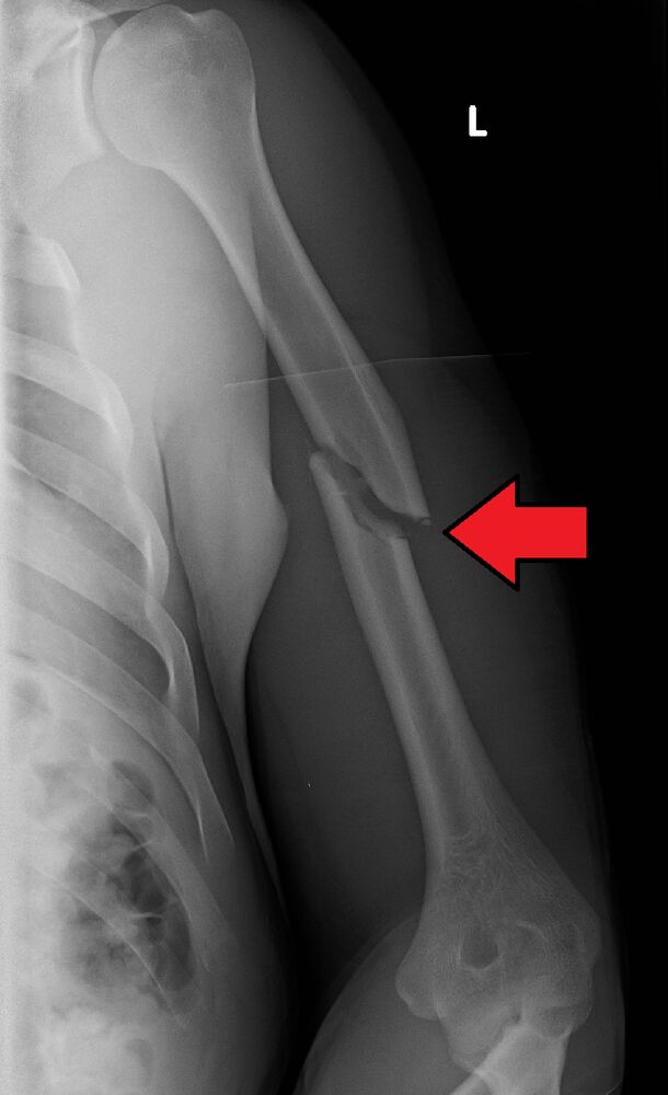
AO/OTA 12A1Spiral, distal third (Holstein–Lewis)
See it in 3D on the real bones, displace and reduce it, with simulated X-ray and CT — free, in your browser.
Open in 3D →The radial nerve lies in the spiral groove on the back of the humerus and pierces the lateral intermuscular septum about 10 cm above the elbow. A spiral fracture of the distal third (Holstein–Lewis) can trap or tear it.
Mechanism
Fall or twisting injury (spiral, e.g. arm wrestling, throwing); direct blow (transverse).
Clinical findings
- Before and after any manipulation test the radial nerve: wrist and finger extension, sensation of the first dorsal web space (wrist drop in about 10%, mostly neurapraxia).
Imaging
- AP and lateral of the whole humerus, including shoulder and elbow.
- Document radial nerve function before and after any manipulation.
- Full-length AP and lateral including shoulder and elbow.
Classification
- 12A
- Simple: A1 spiral, A2 oblique, A3 transverse.
- 12B
- Wedge.
- 12C
- Multifragmentary.
Types shown in 3D
- 12A1
- Simple spiral (here distal third: Holstein–Lewis, radial nerve at risk).
- 12A2
- Simple oblique.
- 12A3
- Simple transverse: highest non-union rate with a brace.
- 12B2
- Wedge (butterfly) fragment.
- 12C2
- Segmental.
Treatment
- Non-operative (most)
- Coaptation (U) splint, then a functional (Sarmiento) brace for 8–12 weeks. Acceptable: < 20° anterior angulation, < 30° varus, < 3 cm shortening. Union in about 90%.
- Operative
- Open fractures, vascular injury, floating elbow, polytrauma, pathological fractures, failure of bracing or non-union: plate (anterolateral or posterior approach, protect the radial nerve) or intramedullary nail. Radial nerve palsy alone is not an indication; palsy appearing after manipulation is debated.
Aftercare
Pendulum exercises; avoid shoulder abduction in early bracing (varus).
Complications
- Radial nerve palsy (recovers in > 70%)
- Non-union (transverse, proximal third, obesity)
- Malunion (usually well tolerated)
See it in 3D on the real bones, displace and reduce it, with simulated X-ray and CT — free, in your browser.
Open in 3D →