 OpenDissect
All fractures › Hip & femur
Pertrochanteric fracture
AO/OTA 31A1–A2Stable and unstable trochanteric fractures
See it in 3D on the real bones, displace and reduce it, with simulated X-ray and CT — free, in your browser.
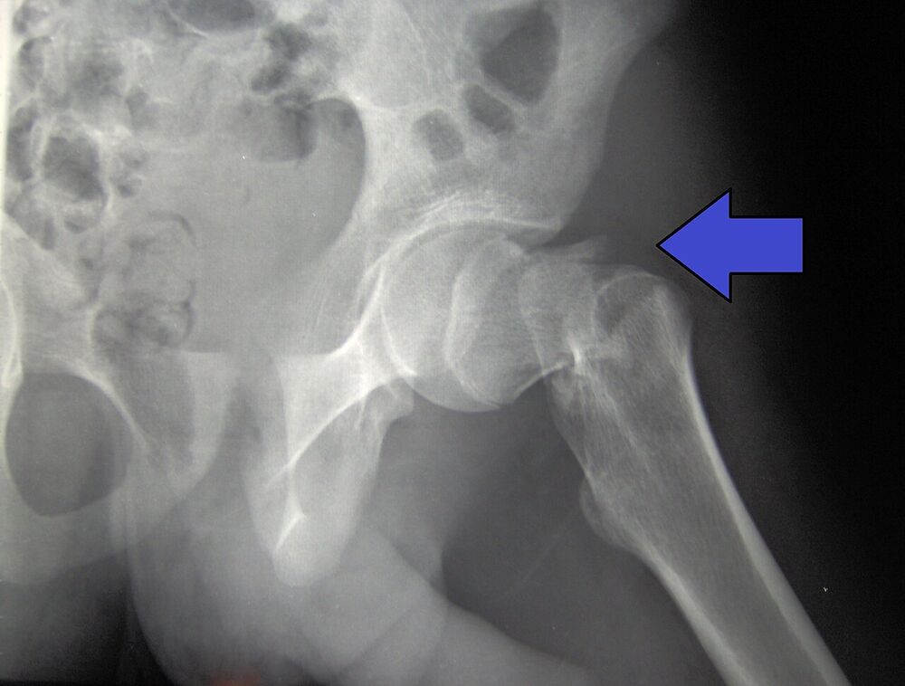
Open in 3D →A fracture running from the greater to the lesser trochanter, outside the capsule. The bone here is well vascularised, so it heals well; the problem is mechanical stability, which depends on the lesser trochanter and the lateral wall.
Mechanism
Fall from standing height in elderly osteoporotic patients (the commonest hip fracture in the very old).
Clinical findings
- Cannot stand, groin and lateral hip pain, bruising over the trochanter.
- Shortened, markedly externally rotated leg (more than in neck fractures).
- Can bleed 1–2 units into the thigh: check haemoglobin.
Imaging
- AP pelvis and AP of the hip.
- Axial view (cross-table lateral): posterior comminution and the lateral wall.
- Full-length femur if a long nail is planned or the shaft is involved. CT for complex or unclear patterns.
- AP pelvis and lateral hip. Look at the lesser trochanter and the lateral wall: if they are broken, the fracture is unstable.
- A traction view in theatre often clarifies the pattern.
Classification
- 31A1
- Simple pertrochanteric: two parts, lateral wall intact (> 20.5 mm). Stable.
- 31A2
- Multifragmentary pertrochanteric with incompetent lateral wall (≤ 20.5 mm). Unstable.
- 31A3
- Intertrochanteric: reverse oblique or transverse line. Very unstable (see its own card).
Types shown in 3D
- 31A1
- Simple pertrochanteric: two parts, lateral wall intact (thicker than 20.5 mm). Stable: sliding hip screw or short nail.
- 31A2.2
- Multifragmentary with one intermediate fragment, usually the lesser trochanter pulled up by iliopsoas. The posteromedial support is lost: unstable, nail preferred.
- 31A2.3
- Multifragmentary with two or more intermediate fragments and an incompetent lateral wall (20.5 mm or less). The shaft medialises: cephalomedullary nail.
Treatment
- Stable (31A1)
- Sliding hip screw (DHS) or a short cephalomedullary nail. The lag screw must sit central-central or slightly inferior in the head with a tip–apex distance under 25 mm.
- Unstable (31A2)
- Cephalomedullary nail (e.g. Gamma or PFNA). The nail acts as a buttress for the lateral wall and prevents the shaft from sliding medially.
- Non-operative
- Only for patients who cannot survive anaesthesia; analgesia, early sitting, accept malunion.
Aftercare
Surgery within 24–48 hours, full weight bearing the next day, orthogeriatric care, fall and osteoporosis assessment.
Complications
- Cut-out of the lag screw (most common failure; tip–apex distance > 25 mm, varus reduction)
- Excessive collapse and shortening
- Peri-implant fracture at the tip of a short nail
- Infection
See it in 3D on the real bones, displace and reduce it, with simulated X-ray and CT — free, in your browser.
Open in 3D →