 OpenDissect
All fractures › Hip & femur
Femoral neck fracture
AO/OTA 31BGarden IV · displaced subcapital
See it in 3D on the real bones, displace and reduce it, with simulated X-ray and CT — free, in your browser.
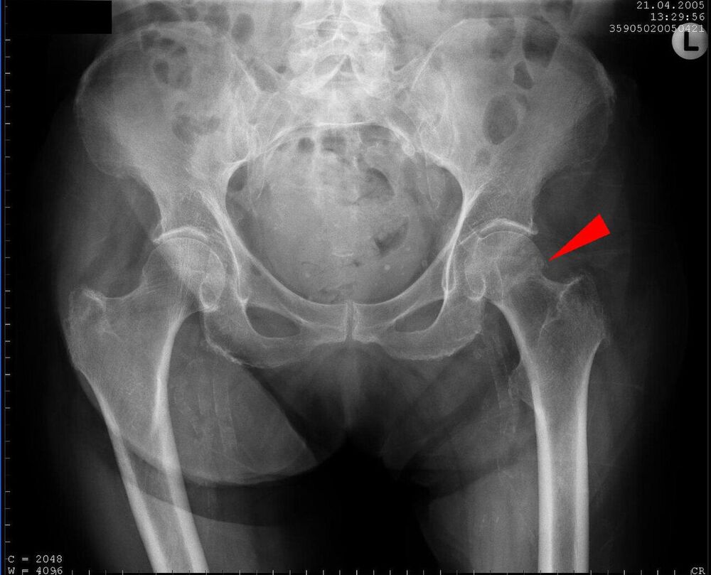
Open in 3D →A break inside the hip capsule, between the femoral head and the trochanters. Typical of older people after a simple fall; in the young it needs high energy. The retinacular vessels that feed the head run along the neck, so displacement can kill the femoral head.
Mechanism
Low-energy fall onto the side of the hip in osteoporotic bone; high-energy axial load in young adults (often with a shaft fracture on the same side).
Clinical findings
- Pain in the groin, cannot bear weight (undisplaced fractures sometimes still walk).
- Displaced: leg shortened and externally rotated.
- Pain on log-rolling and on axial pressure on the heel.
- Look for the cause of the fall (syncope, stroke, medication) and assess cognition and frailty.
Imaging
- AP pelvis and AP of the hip, in slight internal rotation (shows the full length of the neck).
- Axial view of the hip: cross-table lateral (horizontal beam).
- Pain but normal X-rays: MRI (most sensitive), or CT if MRI is not available. About 1 in 10 hip fractures is occult on the first films.
- AP pelvis plus AP and cross-table lateral of the hip.
- Look for a break in the trabecular lines, a step in the cortex and loss of the smooth curve of the neck (Shenton line).
- If the X-ray is normal but the history fits: MRI (best) or CT. A missed undisplaced fracture can displace later.
Classification
- 31B1
- Subcapital (just below the head).
- 31B2
- Transcervical (mid-neck).
- 31B3
- Basicervical (at the base of the neck; behaves more like a trochanteric fracture).
- Garden I–II
- Undisplaced or valgus-impacted. Head blood supply usually preserved.
- Garden III–IV
- Displaced. High risk of avascular necrosis and non-union.
- Pauwels
- Angle of the fracture line to the horizontal: I < 30°, II 30–50°, III > 50°. The steeper the line, the higher the shear and the risk of failure (young patients).
Types shown in 3D
- 31B1
- Subcapital: just below the head.
- 31B2
- Transcervical: through the middle of the neck.
- 31B3
- Basicervical: at the base of the neck. Behaves like a trochanteric fracture (fixed with a sliding hip screw or nail).
- Garden I
- Incomplete or valgus-impacted. Stable; the head blood supply is usually preserved. Fix in situ (or treat as displaced in the very frail).
- Garden II
- Complete but undisplaced: the trabeculae still line up.
- Garden III
- Complete, partially displaced in varus: the trabecular lines of head and acetabulum no longer match.
- Garden IV
- Completely displaced: the head turns back into line with the acetabulum while the shaft rides up and rotates out. Highest risk of avascular necrosis.
- Pauwels I
- Fracture line less than 30° from the horizontal: the load compresses the fracture.
- Pauwels II
- 30–50°: compression and some shear.
- Pauwels III
- More than 50° (vertical): shear dominates and the shaft slides up. High failure rate of fixation in young patients; consider a fixed-angle device.
Treatment
- Undisplaced (Garden I–II)
- Internal fixation in situ: three parallel cannulated screws in an inverted triangle, or a sliding hip screw. In the very old and frail, many surgeons prefer a hemiarthroplasty to avoid a second operation.
- Displaced, older patient
- Replace rather than fix. Hemiarthroplasty for low-demand or cognitively impaired patients; total hip arthroplasty for independent, active patients who walk well.
- Displaced, young patient (< 60)
- Emergency anatomical reduction (closed or open) and stable fixation to save the head: cannulated screws, a sliding hip screw or a fixed-angle device for vertical (Pauwels III) fractures.
- Non-operative
- Rarely: bed-bound patients with very limited life expectancy, after shared decision-making. Even then, surgery is usually the best pain relief.
Aftercare
Operate within 24–48 hours. Full weight bearing after arthroplasty and usually after fixation in the elderly. Orthogeriatric care, delirium prevention, thromboprophylaxis and osteoporosis treatment.
Complications
- Avascular necrosis of the femoral head
- Non-union (especially young, vertical fractures)
- Dislocation after arthroplasty
- Periprosthetic fracture
- High 1-year mortality (about 1 in 4 in the elderly)
See it in 3D on the real bones, displace and reduce it, with simulated X-ray and CT — free, in your browser.
Open in 3D →