 OpenDissect
All fractures › Hip & femur
Femoral shaft fracture
AO/OTA 32A3Simple transverse, midshaft
See it in 3D on the real bones, displace and reduce it, with simulated X-ray and CT — free, in your browser.
Open in 3D →A high-energy injury of the strongest bone of the body. The muscles pull the fragments into a typical deformity: the proximal fragment flexes (iliopsoas), abducts (gluteals) and rotates externally, while the adductors pull the distal fragment medially and the thigh muscles shorten it.
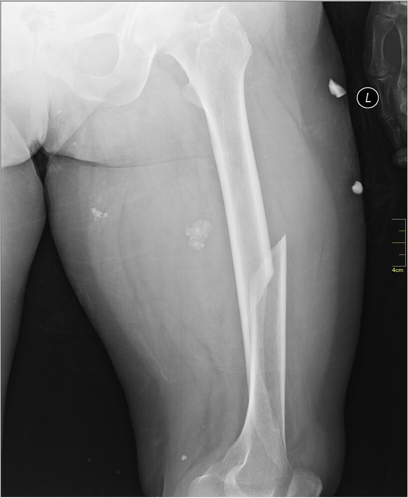
Mechanism
Road traffic accidents, falls from height; low-energy in osteoporotic or pathological bone (look for metastases and atypical fractures on bisphosphonates).
Clinical findings
- Deformed, short, swollen thigh. Can lose 1–1.5 litres of blood.
- ATLS: this is a marker of high energy; look for other injuries (ipsilateral femoral neck fracture in up to 9%).
- Check pulses and the sciatic nerve; compartment syndrome of the thigh is rare but possible.
Imaging
- AP and lateral of the whole femur, including hip and knee.
- Dedicated AP and axial of the hip: an ipsilateral femoral neck fracture is missed in up to a third of cases (CT of the neck in high-energy injuries).
- Trauma CT in polytrauma.
- Full-length AP and lateral of the femur including hip and knee.
- Dedicated hip views or CT to exclude an ipsilateral femoral neck fracture.
Classification
- 32A
- Simple: A1 spiral, A2 oblique (≥ 30°), A3 transverse (< 30°).
- 32B
- Wedge: intact or fragmented wedge; the main fragments touch after reduction.
- 32C
- Multifragmentary: no contact between the main fragments after reduction.
Types shown in 3D
- 32A1
- Simple spiral (torsion). Long fracture surface: heals well, but rotation must be checked.
- 32A2
- Simple oblique (30° or more): the fragments slide and shorten.
- 32A3
- Simple transverse (less than 30°): typical of a direct blow; stable once reduced on a nail.
- 32B2
- Wedge (butterfly) fragment: after reduction the main fragments touch.
- 32C2
- Segmental: a free intermediate segment. No contact between the main fragments; bridge with a nail.
Treatment
- Treatment of choice
- Reamed, locked intramedullary nail (antegrade through the piriformis fossa or the tip of the greater trochanter; retrograde for obesity, ipsilateral knee or acetabular injury, pregnancy or bilateral fractures). Within 24 hours if the patient is stable.
- Damage control
- In the unstable polytrauma patient: temporary external fixator, then convert to a nail when physiology allows (usually within 2 weeks).
- Children
- Under 6 months: Pavlik harness; 6 months – 5 years: hip spica cast; 5–11 years: elastic stable intramedullary nails; adolescents: rigid nail or plate.
Aftercare
Early mobilisation, weight bearing as tolerated after a locked nail in most patterns.
Complications
- Fat embolism and ARDS
- Malrotation (compare with the other side)
- Non-union
- Missed femoral neck fracture
- Pudendal nerve palsy from the traction post
See it in 3D on the real bones, displace and reduce it, with simulated X-ray and CT — free, in your browser.
Open in 3D →