 OpenDissect
All fractures › Hand
Phalangeal fractures
AO/OTA 78Shaft, condylar, bony mallet, volar plate
See it in 3D on the real bones, displace and reduce it, with simulated X-ray and CT — free, in your browser.
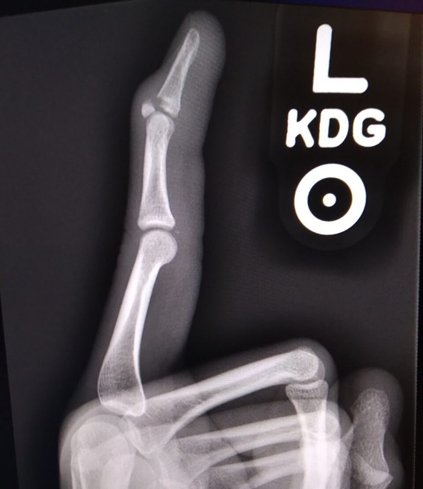
Open in 3D →The commonest fractures of the hand. Tendons lie directly on the phalanges, so displacement, callus and long immobilisation all end up as stiffness. In proximal phalanx shaft fractures the interossei flex the proximal fragment and the central slip extends the distal one: the fracture angulates with its apex volar and shortens the extensor mechanism.
Mechanism
Direct blows, crush, twisting and ball injuries; axial load on the fingertip (bony mallet, condylar fractures); hyperextension of the PIP joint (volar plate avulsion).
Clinical findings
- Swelling, deformity, tenderness; check rotation with the fingers flexed (scissoring) and the alignment of the nails.
- Mallet: the fingertip droops and cannot be actively extended at the DIP joint.
- Volar plate injury: tender over the volar PIP joint after a hyperextension ("jammed finger"); check that the joint is congruent and stable.
- Look for nail-bed lacerations (open tuft fractures).
Imaging
- PA, true lateral and oblique of the injured finger, centred on it.
- The true lateral decides: angulation, joint subluxation (V sign at the PIP joint) and fragment size.
- PA, true lateral and oblique views of the injured finger (not of the whole hand): the lateral shows angulation, subluxation and the size of articular fragments.
- A true lateral of the PIP joint separates a stable small avulsion from an unstable dorsal fracture-dislocation (V sign).
Classification
- 78.x.x.x
- AO/OTA: 78, the finger (1–5), the phalanx (1 proximal, 2 middle, 3 distal) and the segment (1 base, 2 shaft, 3 head); A extra-, B partial, C complete articular.
- Shaft
- Transverse, oblique/spiral, multifragmentary. Proximal phalanx: apex volar. Middle phalanx: apex volar if distal to the FDS insertion, apex dorsal if proximal to it.
- Condylar
- Unicondylar (Weiss–Hastings I–IV) or bicondylar fractures of the head: prone to displace in a splint, so check weekly or fix.
- Bony mallet
- Dorsal base fragment of the distal phalanx with the terminal tendon (small flake Doyle I, articular fragment Doyle IV); significant when > 30–50% of the joint or the distal phalanx subluxes volarly.
- Volar plate / PIP
- Small volar base avulsion of the middle phalanx with a congruent joint (stable) up to the unstable dorsal fracture-dislocation (stable < 30%, tenuous 30–50%, unstable > 50% of the articular surface; Kiefhaber–Stern/Hastings).
Types shown in 3D
- Proximal phalanx shaft (78.2.1.2A)
- The interossei flex the proximal fragment and the central slip extends the distal one: apex volar angulation, which shortens the extensor and gives a PIP extensor lag. Unstable fractures: K-wires, lag screws or a plate.
- Middle phalanx shaft (78.2.2.2A)
- Distal to the FDS insertion the proximal fragment is flexed by the superficial flexor: apex volar. Proximal to it the central slip extends the proximal fragment: apex dorsal.
- Condylar (78.3.1.3B)
- Unicondylar fracture of the proximal phalanx head: the condyle slides proximally and the finger deviates sideways. Prone to displace even when undisplaced: splint with weekly X-rays or fix; displaced fractures need a lag screw or K-wires.
- Bony mallet (78.3.3.1B)
- The terminal extensor tendon pulls off a dorsal base fragment; the distal phalanx drops into flexion. If the phalanx subluxes volarly (or the fragment is more than about a third of the joint) the joint is fixed, usually by extension-block pinning.
- Volar plate avulsion (78.5.2.1B)
- Hyperextension of the PIP joint pulls a small volar base fragment off the middle phalanx with the volar plate. A small fragment with a congruent joint is stable (buddy strapping, early motion); a large one lets the middle phalanx sublux dorsally.
Treatment
- Non-operative
- Stable undisplaced or easily reduced extra-articular fractures: buddy strapping or a splint in the intrinsic-plus position for about 3 weeks, then free motion. Bony mallet with a congruent joint: extension splint of the DIP joint full-time for 6–8 weeks. Small volar plate avulsion: buddy strapping or an extension-block splint and early motion.
- Operative
- Rotation or angulation that cannot be held, unstable spiral fractures, displaced condylar fractures and subluxed joints: K-wires, lag screws (minifragment), plates (shaft, multifragmentary), extension-block pinning for a subluxed bony mallet, and for unstable PIP fracture-dislocations dynamic external fixation, volar plate arthroplasty or hemi-hamate reconstruction.
Aftercare
Start motion as soon as the fixation or the fracture allows (usually by 3 weeks); radiological union lags behind clinical union.
Complications
- Stiffness, PIP flexion contracture
- Extensor lag, swan-neck deformity after mallet injuries
- Rotational malunion
- Post-traumatic arthritis of the PIP or DIP joint
- Nail deformity
See it in 3D on the real bones, displace and reduce it, with simulated X-ray and CT — free, in your browser.
Open in 3D →