 OpenDissect
All fractures › Hand
Metacarpal fractures
AO/OTA 77Boxer's, shaft, CMC fracture-dislocation
See it in 3D on the real bones, displace and reduce it, with simulated X-ray and CT — free, in your browser.
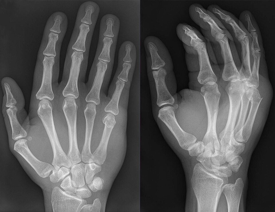
Open in 3D →Metacarpal fractures make up roughly a third of hand fractures, most of them in the ring and little rays. The intrinsic muscles flex the distal fragment, so neck and shaft fractures angulate with the apex dorsal. The deformity that matters most is rotation: a few degrees at the fracture become a finger that crosses its neighbour (scissoring) when the fist is closed.
Mechanism
Punching a hard object with a closed fist (neck of the fifth, "boxer's fracture"); twisting injuries (spiral shaft fractures); direct blows and crush (transverse and multiple fractures); axial load on a flexed metacarpal (base and carpometacarpal fracture-dislocations).
Clinical findings
- Swelling and loss of the knuckle contour; tenderness over the fracture.
- Rotation: ask the patient to make a gentle fist – all fingertips should point to the scaphoid tubercle and the nail plates should line up. Compare with the other hand.
- Lacerations over the knuckles after a punch are human bites until proven otherwise ("fight bite"): explore, wash out and give antibiotics.
- Extensor lag (pseudoclawing) after neck fractures; check the extensor tendons.
Imaging
- PA, lateral and oblique views of the hand.
- Lateral: measure the angulation of the neck or shaft. Pronated oblique (about 30°) for the 4th and 5th CMC joints; CT if a CMC fracture-dislocation is suspected.
- Rotation is a clinical diagnosis: examine the fist.
- PA, lateral and oblique views of the hand. On the lateral, measure the angle of the neck against the shaft.
- For the fourth and fifth CMC joints a 30° pronated oblique view (and, if in doubt, CT): the dislocation is easy to miss on the PA.
Classification
- 77.x
- AO/OTA: bone 77, then the ray (1 thumb to 5 little finger), then the segment: .1 proximal end (base), .2 shaft, .3 distal end (neck and head). A extra-articular, B partial articular, C complete articular.
- Neck (boxer's)
- Apex dorsal angulation of the head. Accepted angulation increases from radial to ulnar because the 4th and 5th CMC joints are mobile: roughly < 15° for the index, < 20° middle, < 30–40° ring, < 50–70° little finger.
- Shaft
- Transverse (apex dorsal angulation), oblique/spiral (shortening and rotation), multifragmentary. Shortening of a few millimetres is well tolerated; rotation is not.
- Base / CMC
- Fracture-dislocations of the 4th and 5th CMC joints (the 5th base is pulled proximally and ulnarly by the extensor carpi ulnaris: "reverse Bennett").
- Head
- Intra-articular, often comminuted; may be a fight bite.
Types shown in 3D
- Neck of the 5th (boxer's, 77.5.3A)
- The commonest metacarpal fracture: the head tips into the palm (apex dorsal angulation), pulled by the interossei, with volar comminution. The little finger tolerates a lot of angulation thanks to its mobile CMC joint, but no rotation.
- Shaft, transverse (77.2.2A)
- Direct blow: the distal fragment is flexed by the intrinsics (apex dorsal). Less angulation is accepted than at the neck (about 10° index and middle, 20–30° ring and little).
- Shaft, spiral (77.4.2A)
- Twisting injury: the long oblique fracture lets the distal fragment shorten and rotate. A few degrees of rotation at the shaft become a finger that overlaps its neighbour in a fist (scissoring): this is the main indication for surgery (lag screws or plate).
- Base, 4th–5th CMC fracture-dislocation (77.5.1B)
- Axial load on the flexed ring and little metacarpals: the bases dislocate dorsally off the hamate, the 5th with a small radial base fragment that stays in place ("reverse Bennett"); extensor carpi ulnaris pulls the shaft proximally. Easily missed: look at the CMC joints on a pronated oblique view.
Treatment
- Non-operative
- Most neck and shaft fractures: closed reduction if needed (Jahss manoeuvre for the neck), then a functional splint, buddy strapping or a cast in the intrinsic-plus position (MCP joints flexed 70–90°, IP joints straight) for 3–4 weeks. Many little-finger neck fractures are treated with early motion and no reduction at all.
- Operative
- Any rotational malalignment, angulation beyond the accepted limits, shortening > 5 mm, multiple metacarpal fractures, open fractures and displaced intra-articular fractures. Options: K-wires (antegrade "bouquet" pinning or transverse pins into the neighbouring metacarpal), intramedullary headless screws, lag screws for long spiral fractures, plates for multifragmentary or multiple fractures.
- CMC fracture-dislocation
- Closed reduction and percutaneous pinning (to the hamate or the neighbouring metacarpal), or open reduction if the joint surface is displaced or the reduction cannot be held.
Aftercare
Early active finger motion is the priority. Union is usually clinical by 4–6 weeks; contact sport after about 6–8 weeks.
Complications
- Rotational malunion (scissoring)
- Extensor lag, loss of knuckle prominence
- Stiffness of the MCP joints (immobilised in extension)
- Infection after a fight bite
- Post-traumatic CMC arthritis
See it in 3D on the real bones, displace and reduce it, with simulated X-ray and CT — free, in your browser.
Open in 3D →