 OpenDissect
All fractures › Elbow, forearm & wrist
Olecranon fracture
AO/OTA 2U1BSimple transverse, displaced
See it in 3D on the real bones, displace and reduce it, with simulated X-ray and CT — free, in your browser.
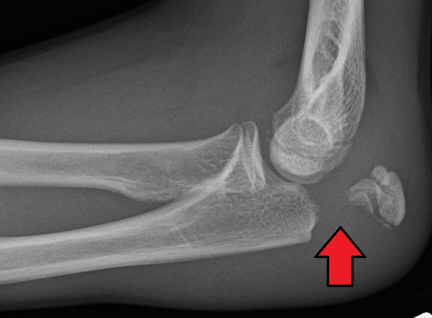
Open in 3D →The triceps inserts on the olecranon, so a transverse fracture is pulled apart and the patient loses active elbow extension. It is an intra-articular fracture of the trochlear notch.
Mechanism
Fall onto the point of the elbow (direct) or onto the outstretched hand with a contracting triceps.
Clinical findings
- Palpable gap, inability to extend the elbow against gravity. Check the ulnar nerve.
Imaging
- AP and true lateral of the elbow (the lateral shows displacement and comminution).
- Look for an associated radial head fracture or dislocation.
- A true lateral view shows displacement and comminution; look for an associated radial head fracture or dislocation (fracture-dislocation).
Classification
- Mayo I
- Undisplaced.
- Mayo II
- Displaced, stable elbow (A non-comminuted, B comminuted).
- Mayo III
- Displaced, unstable elbow.
Types shown in 3D
- Mayo I
- Undisplaced (< 2 mm), extensor mechanism intact: early protected motion.
- Mayo IIA
- Displaced, simple, stable elbow: tension-band wiring.
- Mayo IIB
- Displaced, comminuted, stable elbow: plate fixation (tension band would collapse the notch).
- Mayo III
- Displaced and unstable: fracture-dislocation (the forearm slips forward with the distal ulna). Plate and restore the trochlear notch; check the coronoid and radial head.
Treatment
- Non-operative
- Undisplaced with intact extension, or very low-demand elderly patients (often good function despite displacement): sling, early motion.
- Operative
- Tension-band wiring for simple transverse fractures; a contoured posterior locking plate for oblique, comminuted fractures and fracture-dislocations.
Aftercare
Early active motion after stable fixation.
Complications
- Symptomatic hardware (frequent)
- Loss of extension
- Ulnar neuritis
- Post-traumatic arthritis
See it in 3D on the real bones, displace and reduce it, with simulated X-ray and CT — free, in your browser.
Open in 3D →