 OpenDissect
All fractures › Elbow, forearm & wrist
Monteggia & Galeazzi injuries
AO/OTA 2U2 / 2R2Forearm fracture with a dislocated joint
See it in 3D on the real bones, displace and reduce it, with simulated X-ray and CT — free, in your browser.
Open in 3D →The radius and ulna form a ring with two joints. If one bone is broken and shortened, the other must dislocate at one end. Monteggia: ulnar fracture with dislocation of the radial head. Galeazzi: radial shaft fracture (distal third) with dislocation of the distal radioulnar joint (DRUJ).
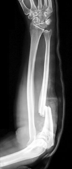
Mechanism
Falls on the outstretched hand with forced pronation; direct blows ("nightstick" mechanism).
Clinical findings
- Any isolated ulnar fracture: check the radial head. Any radial shaft fracture: check the DRUJ (prominent ulnar head, painful rotation).
- Monteggia: posterior interosseous nerve palsy (finger drop).
Imaging
- AP and lateral of the whole forearm, including elbow and wrist: always image the joint above and below a forearm fracture.
- Radiocapitellar line on every elbow view (Monteggia); distal radioulnar joint on the wrist lateral (Galeazzi).
- Always X-ray the whole forearm including elbow and wrist. The radiocapitellar line must point at the capitellum in every view.
Classification
- Bado I–IV
- Monteggia by the direction of the radial head dislocation: I anterior (most common), II posterior, III lateral, IV with radial shaft fracture.
- Galeazzi
- Radial shaft fracture with DRUJ injury; fractures within 7.5 cm of the wrist are more often unstable.
Types shown in 3D
- Bado I
- Ulnar shaft fracture angulated anteriorly with anterior dislocation of the radial head (most common, especially in children).
- Bado II
- Posterior angulation and posterior dislocation of the radial head (adults, often with a radial head fracture).
- Bado III
- Ulnar metaphyseal fracture with lateral dislocation of the radial head (children).
- Bado IV
- Fracture of both bones at the same level with anterior dislocation of the radial head.
- Galeazzi
- Radial shaft fracture in the distal third with dislocation of the distal radioulnar joint: the radius shortens and the ulnar head becomes prominent. Plate the radius, then test the DRUJ.
Treatment
- Adults
- Open reduction and plate fixation of the broken bone restores length and usually reduces the dislocated joint. Then test the joint: an unstable DRUJ is pinned or held in supination; a radial head that stays dislocated suggests malreduction of the ulna or interposed tissue.
- Children
- Monteggia: closed reduction of the ulna (plastic deformation counts!) and above-elbow cast; unstable patterns get an intramedullary wire or plate.
Aftercare
Early motion after plating.
Complications
- Missed dislocation (chronic Monteggia is difficult to treat)
- Loss of forearm rotation
- Nerve palsy
See it in 3D on the real bones, displace and reduce it, with simulated X-ray and CT — free, in your browser.
Open in 3D →