 OpenDissect
All fractures › Chest wall
Sternal fractures
Chest wallTransverse body fracture, manubriosternal dislocation
See it in 3D on the real bones, displace and reduce it, with simulated X-ray and CT — free, in your browser.
Open in 3D →Most sternal fractures are transverse fractures of the body or at the manubriosternal junction, from a direct blow to the front of the chest. In isolation they are usually benign, but they mark a high-energy impact: look for blunt cardiac injury, thoracic spine fractures and other chest injuries.
Mechanism
Steering wheel or seatbelt in frontal collisions, direct blows; flexion–compression injuries of the thoracic spine (indirect fracture); insufficiency fractures in osteoporotic, kyphotic patients.
Clinical findings
- Anterior chest pain, tenderness, sometimes a palpable step or crepitus.
- Screen for blunt cardiac injury: 12-lead ECG and troponin on arrival; normal both means significant cardiac injury is very unlikely. Echocardiography for arrhythmia, haemodynamic instability or abnormal results.
- Examine and image the thoracic spine: a displaced sternal fracture with a flexion mechanism is often paired with a thoracic spine fracture.
Imaging
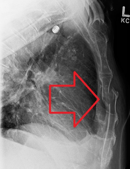
- Lateral sternal view (the frontal chest film usually misses it).
- CT chest with sagittal reformats in major trauma.
- 12-lead ECG and troponin for blunt cardiac injury; echocardiography if abnormal or unstable.
- Lateral sternal view: the key radiograph (the fracture is often invisible on the frontal chest X-ray).
- CT chest with sagittal reformats in major trauma; look for retrosternal haematoma and mediastinal injury (CT angiography if suspected).
- Ultrasound can show a cortical step at the bedside.
Classification
- Location
- Manubrium, manubriosternal junction, body (most common), xiphoid.
- Displacement
- Undisplaced, or displaced with overlap of the fragments (usually the lower fragment in front of the upper).
- Manubriosternal dislocation
- Type 1: body displaced behind the manubrium (direct blow). Type 2: body in front of the manubrium (indirect, flexion–compression with a thoracic spine fracture).
Types shown in 3D
- Undisplaced transverse (body)
- The commonest pattern: a transverse line through the upper body. Check ECG and troponin; analgesia.
- Displaced transverse (body)
- The lower fragment is displaced forwards relative to the upper one. Look for a thoracic spine fracture and retrosternal haematoma; plate fixation if unstable or very painful.
- Manubriosternal dislocation (type 1)
- The body is driven behind the manubrium by a direct blow. Usually reduced and fixed if displaced; rule out mediastinal injury.
Treatment
- Non-operative (most)
- Analgesia, breathing exercises; patients with an isolated undisplaced fracture and normal ECG and troponin can usually go home.
- Operative
- Severe displacement with instability or deformity, intractable pain, respiratory compromise, an unstable "sternal flail", or painful non-union: open reduction and anterior locking plate fixation.
Aftercare
Symptoms settle over 6–12 weeks; no lifting > 5 kg and no contact sports for about 6 weeks after fixation.
Complications
- Blunt cardiac injury (arrhythmia, contusion)
- Non-union or painful malunion
- Missed thoracic spine fracture
- Retrosternal haematoma, rarely mediastinal vascular injury
See it in 3D on the real bones, displace and reduce it, with simulated X-ray and CT — free, in your browser.
Open in 3D →