 OpenDissect
All fractures › Chest wall
Rib fractures and flail chest
Chest wall (CWIS)Serial fractures, flail segment, SSRF
See it in 3D on the real bones, displace and reduce it, with simulated X-ray and CT — free, in your browser.
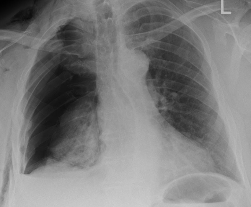
Open in 3D →The most common injury after blunt chest trauma. The danger lies less in the bone than in what comes with it: pneumothorax, haemothorax, pulmonary contusion, and pain that stops the patient from breathing deeply and coughing. In older patients each additional broken rib increases the risk of pneumonia and death. Three or more adjacent ribs broken in two places make a flail segment that moves paradoxically with breathing.
Mechanism
Direct blows, road traffic accidents and falls; simple falls in the elderly. First and second rib fractures need high energy. Lower ribs (9–12) overlie the liver, spleen and kidneys.
Clinical findings
- Point tenderness, pain on breathing and coughing, crepitus; paradoxical movement of a flail segment (may be masked by splinting or ventilation).
- Primary survey: tension pneumothorax, massive haemothorax, open chest wound; oxygen saturation and respiratory rate.
- Quantify pain and respiratory function (spirometry, cough); older patients and those with ≥ 3 fractures belong in a monitored setting.
Imaging
- Chest X-ray (erect PA if possible; supine AP in the trauma bay) for pneumothorax, haemothorax and contusion.
- CT chest in major trauma or when several fractures, flail chest or associated injuries are suspected.
- Ultrasound (eFAST) for pneumothorax and haemothorax at the bedside. Dedicated rib series rarely change management.
- Chest X-ray: pneumothorax, haemothorax, contusion; it misses a large share of rib fractures.
- CT chest is the standard in major trauma: number, location and displacement of fractures, flail segments, contusion and associated injuries.
- Ultrasound (eFAST) for pneumothorax and haemothorax at the bedside. Upper rib fractures: think of aortic and subclavian injury (CT angiography); lower ribs: liver, spleen and kidney.
Classification
- CWIS taxonomy
- Each fracture by type (simple, wedge, complex), displacement (undisplaced, offset, displaced with no cortical contact) and location (anterior, lateral, posterior sectors).
- Flail segment
- Three or more consecutive ribs each broken in two or more places; flail chest = the segment moves paradoxically.
- Associated
- Pneumothorax, haemothorax, pulmonary contusion, injury to the great vessels (ribs 1–2), liver, spleen and kidney (ribs 9–12).
Types shown in 3D
- Single undisplaced
- One lateral rib fracture with cortical contact. Analgesia and breathing exercises; usually heals in about 6 weeks.
- Serial displaced (ribs 4–6)
- Three adjacent ribs broken once each, the anterior fragments displaced inwards. Painful; watch for pneumothorax and haemothorax, and consider SSRF if severely displaced with intractable pain.
- Flail segment (ribs 4–7)
- Four consecutive ribs broken in front and behind: the floating segment is sucked in on inspiration (paradoxical movement), usually over a pulmonary contusion. Analgesia, ventilation if needed, SSRF for respiratory failure.
- Lower ribs (10–11)
- Posterolateral fractures of the lower ribs: the liver lies behind them on the right (spleen on the left), the kidney further back. CT abdomen.
Treatment
- All patients
- Multimodal analgesia (paracetamol, NSAIDs if safe, opioids sparingly), regional techniques for multiple fractures (thoracic epidural, paravertebral, erector spinae or serratus anterior plane block), physiotherapy, incentive spirometry, early mobilisation. Avoid fluid overload with pulmonary contusion.
- Pneumothorax and haemothorax
- Chest drain for pneumothorax and haemothorax; small occult pneumothoraces and small haemothoraces (< about 300 ml) can be observed in stable patients. Thoracotomy for ≥ 1500 ml at insertion, > 200 ml/h for 2–4 h, or ongoing transfusion need; VATS within 3–5 days for retained haemothorax.
- Surgical stabilisation (SSRF)
- Flail chest with respiratory failure or failure to wean; severely displaced fractures with intractable pain, chest wall deformity or volume loss despite good analgesia; symptomatic non-union. Plates or intramedullary splints, ideally within 72 hours. Guidelines suggest SSRF for adult flail chest to reduce mortality, ventilator days and pneumonia.
Aftercare
Pain usually settles over 3–6 weeks; avoid contact sports for 6–8 weeks.
Complications
- Pneumonia and respiratory failure (elderly)
- Retained haemothorax and empyema
- Delayed pneumothorax or haemothorax
- Chronic pain, intercostal neuralgia
- Non-union
See it in 3D on the real bones, displace and reduce it, with simulated X-ray and CT — free, in your browser.
Open in 3D →