 OpenDissect
All fractures › Pelvis & spine
Acetabular fractures
AO/OTA 62Columns and walls (Letournel)
See it in 3D on the real bones, displace and reduce it, with simulated X-ray and CT — free, in your browser.
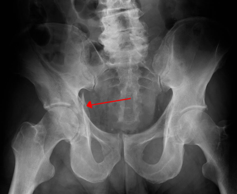
Open in 3D →The acetabulum sits in an inverted Y of two columns: the anterior (iliopectineal) and posterior (ilioischial). Letournel described five elementary and five associated patterns; recognising them on the AP and Judet views guides the approach.
Mechanism
Force transmitted through the femoral head: dashboard injury (posterior wall, often with hip dislocation); falls on the side in the elderly (anterior column, quadrilateral plate).
Clinical findings
- Check the sciatic nerve (posterior wall fractures with dislocation). Reduce a dislocated hip urgently.
Imaging
- AP pelvis.
- Judet views: obturator oblique (anterior column, posterior wall) and iliac oblique (posterior column, anterior wall).
- CT with 3D reconstructions is the standard for classification (Letournel) and planning.
- AP pelvis, Judet views (obturator and iliac oblique) and CT with 3D reconstructions. Six lines on the AP: iliopectineal, ilioischial, teardrop, roof, anterior and posterior wall.
Classification
- Elementary
- Posterior wall, posterior column, anterior wall, anterior column, transverse.
- Associated
- Posterior column + wall, transverse + posterior wall, T-shaped, anterior column + posterior hemitransverse, both columns ("spur sign").
- AO/OTA 62
- 62A one column/wall, 62B transverse-type (part of the roof attached to the ilium), 62C both columns (articular surface detached from the ilium).
Types shown in 3D
- Posterior wall
- The most common: dashboard injury with posterior hip dislocation. Check the sciatic nerve; fix through a Kocher–Langenbeck approach.
- Posterior column
- The ischial part separates from the sciatic notch down through the joint into the obturator ring; the head follows it medially.
- Anterior column
- Typical of falls on the side in the elderly; the anterior part of the joint and the superior ramus separate. Anterior approach (ilioinguinal or modified Stoppa).
- Transverse
- One line across the acetabulum splits the innominate bone into an upper (iliac) and a lower (ischiopubic) half; the head can protrude centrally.
- Both columns
- No part of the joint stays attached to the intact ilium ("floating acetabulum", spur sign on the obturator oblique view). Secondary congruence can allow non-operative care in the elderly.
Treatment
- Non-operative
- Displacement < 2 mm, roof arcs intact, stable hip, or a both-column fracture with secondary congruence: protected weight bearing 6–8 weeks.
- Operative
- Displaced fractures in fit patients: anatomical reduction and plates/screws through the Kocher–Langenbeck (posterior) or ilioinguinal / modified Stoppa (anterior) approaches, within about 2 weeks. Elderly with poor bone and femoral head impaction: consider acute total hip arthroplasty with fixation.
Aftercare
Toe-touch weight bearing 8–12 weeks.
Complications
- Post-traumatic arthritis
- Heterotopic ossification (posterior approach)
- Sciatic nerve injury
- Avascular necrosis of the femoral head
- Thromboembolism
See it in 3D on the real bones, displace and reduce it, with simulated X-ray and CT — free, in your browser.
Open in 3D →